IMAGE

Fig. 2

ID
ZDB-IMAGE-190723-1903
Source
Figures for Felker et al., 2018
Image
Figure Caption

Fig. 2

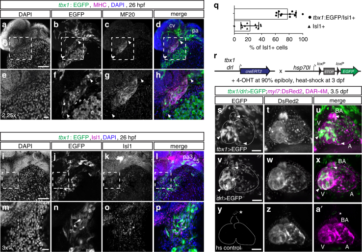
tbx1+ cells contribute to LPM-derived cardiac lineages. ap Representative maximum intensity projections of whole-mount tbx1:EGFP-expressing embryos counterstained for anti-EGFP and anti-MHC (n = 3) (ah) or anti-Isl1 (n = 11) (ip) at 26 hpf; lateral views, anterior to the left. adtbx1 reporter expression can be detected in the MHC-positive linear heart tube and in the MHC-negative poles at the cardiac inflow and outflow tracts (arrowheads); eh depicts a 2.25x magnification of the framed area in ad. tbx1:EGFP also marks the pharyngeal arches (pa) and endothelial cells of the cranial vasculature (cv) (d). iptbx1 reporter-expressing cells at the IFT co-express the SHF marker Isl1 (asterisks n, o); mp depicts a 3x magnification of the framed area in il. q Quantification of tbx1:EGFP/Isl1 double- compared to Isl1 single-positive cells at the IFT of the linear heart tube, n = 11 individual embryos analyzed, means ± s.d. r Lineage tracing of tbx1 and drl reporter-expressing cells, shown in representative embryos. tbx1:creERT2 (n = 11) or drl:creERT2 (n = 3) transgenics, respectively, were crossed to the ubiquitous hsp70l:Switch loxP tracer line, embryos were 4-OHT-induced at 90% epiboly, and heat-shocked at 3 dpf. s-a’ Live SPIM imaging of still hearts of representative lineage-traced and control embryos; maximum intensity projections of ventral views, anterior to the top, dashed outlines mark the heart with the bulbus arteriosus (BA), atrium (A), and ventricle (V). sutbx1:creERT2 lineage tracing (tbx1 > EGFP) at late gastrulation labels myl7:DsRed2-expressing cardiomyocytes (arrowheads) in the ventricle and inflow tract of the atrium, and DAR-4M-stained cells (arrowhead) in the BA. vxdrl:creERT2-mediated lineage tracing (drl > EGFP) at 90% epiboly marks all cardiomyocytes (arrowheads) in the ventricle and atrium, BA cells (arrowhead), and the endocardium (arrows). y-atbx1:creERT2;hsp70l:Switch transgenics without 4-OHT treatment and heat-shocked at 3 dpf show no specific EGFP expression (asterisks mark the autofluorescent pigment cell). Scale bars 20 μm (eh, mp), 100 μm (ad, il, sa’)

Acknowledgments
This image is the copyrighted work of the attributed author or publisher, and ZFIN has permission only to display this image to its users. Additional permissions should be obtained from the applicable author or publisher of the image. Full text @ Nat. Commun.