IMAGE

Fig. 3.

ID
ZDB-IMAGE-190723-1578
Source
Figures for Welker et al., 2016
Image
Figure Caption

Fig. 3.

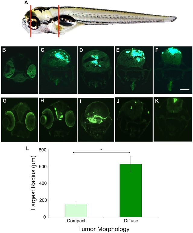
GBM9 tumor cells grow throughout the brain tissue. (A) Representative area sectioned (red lines) in a 7 dpt zebrafish. (B-F) Transverse 20-μm-thick cryosections of a GBM9 compact tumor at the level of the forebrain (B), midbrain (C,D) and hindbrain (E,F). (G-K) Transverse cryosections of a diffuse tumor at the level of the forebrain (G,H) midbrain (I) and hindbrain (J,K). (L) Based on morphology, tumors were scored as compact (light green bar) or diffuse (dark green bar) then measured by Sholl analysis at 7 dpt to quantify cell spread. Largest radius (in micrometers) is the measure of the farthest radius intersecting a cell from the injection site. n=10 per group; 20 animals total. *P<0.001. Scale bar: 100 μm for B-K.

Acknowledgments
This image is the copyrighted work of the attributed author or publisher, and ZFIN has permission only to display this image to its users. Additional permissions should be obtained from the applicable author or publisher of the image. Full text @ Dis. Model. Mech.