IMAGE

Figure 6.

ID
ZDB-IMAGE-190723-1564
Source
Figures for Eckert et al., 2019
Image
Figure Caption

Figure 6.

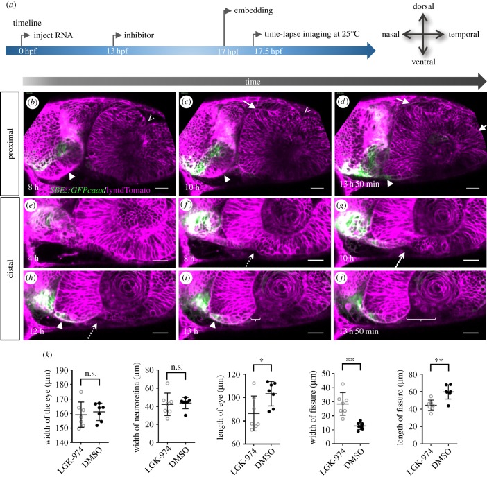
Wnt-signalling inhibition affects optic cup morphogenesis and prevents TGFβ-signalling positive cells from entering the ventral part of the optic cup. (a) Timeline of the experimental procedure, and orientation of the eye. (bj) Four-dimensional dataset of a developing optic cup. TGFβ reporter (green); cell membranes (lyntdTomato, magenta). One optical section in a proximal (bd) and one in a distal region (ej) over time (left to right). TGFβ-signalling-positive cells are located in the misshaped optic stalk/forebrain (arrowhead). Few TGFβ-signalling-positive cells reach the nasal ventral part of the developing optic cup (h,i, arrowhead). The dorsal fissure (bc, marked with v) seems to close over time. Ectopic domains of the presumptive neuroretina can be seen in the lens-averted dorsal domain (cd, arrows). Even though TGFβ-positive cells do not move into the eye, a nasal fissure margin is visible. On the temporal side, the ventral perpendicular flow seems corrupted, affecting the formation of the temporal fissure margin. The distal flow in the ventral domains, both nasal and temporal, seems unaffected resulting in an optic fissure being visible in distal domains (i,j brackets). A dotted arrow indicates where the fissure will open (5 fish in 1 experiment, 3 TGFB reporter, 2 WT). Scale bar, 25 µm. (k) Quantification of distinct morphological parameters of LGK-974-treated embryos versus DMSO-treated embryos. *p < 0.05; **p < 0.005.

Acknowledgments
This image is the copyrighted work of the attributed author or publisher, and ZFIN has permission only to display this image to its users. Additional permissions should be obtained from the applicable author or publisher of the image. Full text @ Open Biol.