IMAGE

Figure 5.

ID
ZDB-IMAGE-190723-1563
Source
Figures for Eckert et al., 2019
Image
Figure Caption

Figure 5.

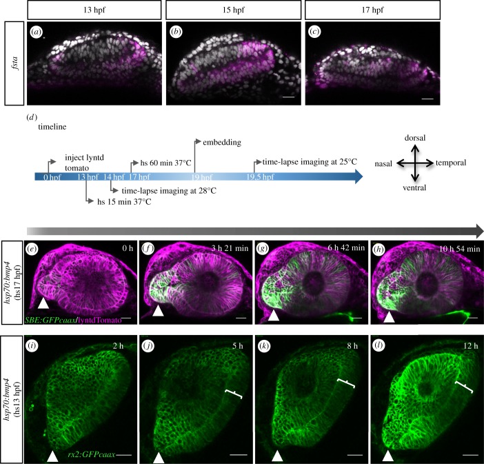
Induced expression of bmp4 hampers optic fissure formation. In situ hybridizations for fsta (ac) for 13, 15 and 17 hpf in WT embryos. fsta is expressed, from temporal to the ventral transition zone to the optic stalk. (d) Timeline of the experimental procedure and orientation of the eye, heat shocks (hs) performed at 17 hpf are displayed on top of the arrow, hs performed at 13 hpf below the arrow. (eh) Lateral view of optic cup development in the tg(hsp70:bmp4) background, visualized by lyntdTomato (mRNA), tg(SBE:GFPcaax). bmp4 induced at 17 hpf hampers proximal optic fissure morphogenesis (10 fish in 1 experiment). The optic stalk is in continuation to the lens-averted domains of the developing optic cup (e, arrowhead). Asterisk marks the optic ventricle. In temporal and dorsal regions, the lens-averted layer is being integrated into the optic cup (f–h). Cells from the lens-averted layer of the optic cup are not properly integrated into the lens-facing domain. The connection of the optic stalk to the lens-averted domain is maintained and the optic fissure is not formed in the proximal domain. The TGFβ signalling activity can be seen in the optic cup, in the absence of orderly tissue dynamics. See figure 4 as control. (il) Lateral view of optic cup development in the tg(hsp70:bmp4) background, visualized by tg(rx2:GFPcaax). bmp4 induced at 13 hpf results in an absence of the optic fissure (4 fish in 1 experiment). The optic stalk is misshaped (arrowhead). On the temporal side, a persisting lens-averted domain is visible (brackets jl). Scale bar, 25 μm. See electronic supplementary material, figure 5 supplement 1 O–X as control.

Acknowledgments
This image is the copyrighted work of the attributed author or publisher, and ZFIN has permission only to display this image to its users. Additional permissions should be obtained from the applicable author or publisher of the image. Full text @ Open Biol.