IMAGE

Figure 4.

ID
ZDB-IMAGE-190723-1562
Source
Figures for Eckert et al., 2019
Image
Figure Caption

Figure 4.

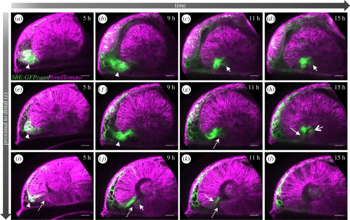
TGFβ-signalling-positive cells are secondarily added to the optic fissure margins. (al) Four-dimensional dataset of the developing optic cup, TGFβ reporter (green), cell membranes (lyntdTomato, magenta). Presented are three different optical planes (top to bottom) over time (left to right). TGFβ reporter activity in the optic stalk (a,e,i, arrowhead). TGFβ-signalling-positive cells move from the optic stalk into the optic cup (ck, arrow) (8 fish in 4 experiments; 3 fish in 2 experiments). The optic stalk is predominantly connected to the nasal optic cup (a,b,e,f). At the end of the flow, TGFβ-signalling-positive cells populate the most proximal domain of the optic fissure (c,d,f,g) (3 fish in 2 experiments). From here, these cells also populate the temporal fissure margin (c,d,f,g,h,j). The optic fissure is marked with a dotted arrow. Lateral view, nasal to the left; scale bar, 25 µm.

Acknowledgments
This image is the copyrighted work of the attributed author or publisher, and ZFIN has permission only to display this image to its users. Additional permissions should be obtained from the applicable author or publisher of the image. Full text @ Open Biol.