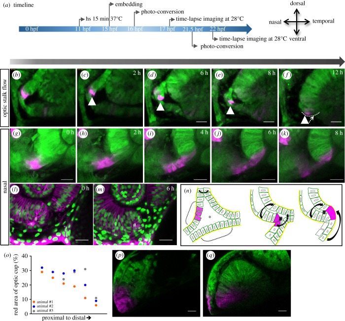
IMAGE

Figure 3.

ID
ZDB-IMAGE-190723-1561
Source
Figures for Eckert et al., 2019
Image
Figure Caption

Figure 3.

Development of the nasal fissure margin. (a) Timeline of experimental procedure and orientation of the eye (timeline for bf displayed on top of the arrow, for gk below). (bf and gk) Close ups of the developing nasal ventral optic cup labelled with tg(HSP70::kaede), photo-converted cells (magenta) derived from the optic stalk translocate from the optic stalk into the nasal fissure margin (12 fish in 6 experiments; not all of them are kaede experiments). Lateral view, nasal to the left, scale bar: 25 µm. (lm) In distal domains, a turbulence or vortex of cells can be appreciated (hash), in this domain, two flow movements collide, one over the distal rim, one from the optic stalk. Cells remaining in the RPE domain flatten and thus obtain RPE cell shape (labelled by tg(bact:H2BGFP) and tg(bact:lyntdTomato)). (n) Scheme of nasal fissure margin development. In proximal regions of the early optic cup, the optic stalk consists of two layers (dashed brackets), which are connected to the lens-averted domains of the optic cup (see also figure 1g) and border the optic ventricle (asterisk). Over time, the upper layer is moving into the optic fissure margin. The distal and optic stalk flow movements are indicated by black arrows. The twist of the optic stalk could easily be driven by the distal flow on the nasal side which is flowing over the optic stalk. Eventually, stalk-derived cells are integrated via the nasal fissure margin. (o) Optic stalk contribution to the ventral neuroretina (stalks of three animals were converted and the red area was measured separately). (p) Optic vesicle from animal #2 before fissure development. Parts of the optic stalk were converted. (q) Optic cup from animal #2 after fissure development. Photo-converted cells moved into the ventral area of the optic cup. Scale bar, 25 μm.

Acknowledgments
This image is the copyrighted work of the attributed author or publisher, and ZFIN has permission only to display this image to its users. Additional permissions should be obtained from the applicable author or publisher of the image. Full text @ Open Biol.