IMAGE

Figure 1.

ID
ZDB-IMAGE-190723-1558
Source
Figures for Eckert et al., 2019
Image
Figure Caption

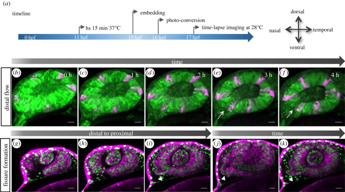
Figure 1.

Transformation of the optic vesicle into the optic cup. (a) Timeline of experimental procedure (for bf) and orientation of the eye. (bf) Distal flow and fissure generation. (b) Photo-converted domains (magenta) in the lens-averted layer move over the distal rims into the lens-facing domain (cf). During this process, the fissure is induced (dotted arrow). (gi) The optic fissure is generated from distal to proximal. (ik) The optic stalk (arrowhead) is connected to the nasal lens-averted domain of the optic vesicle, imaging starts (gj) at 19 hpf; scale bar, 25 µm.

Acknowledgments
This image is the copyrighted work of the attributed author or publisher, and ZFIN has permission only to display this image to its users. Additional permissions should be obtained from the applicable author or publisher of the image. Full text @ Open Biol.