IMAGE

Fig. 1

ID
ZDB-IMAGE-190723-1435
Genes
Source
Figures for Pei et al., 2019
Image
Figure Caption

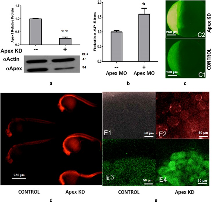
Fig. 1 Loss of Apex1 protein results in increased oxidative damage, AP sites and ROS.

a Western blot analysis of Apex1 knockdown by morpholino (MO). Upper panel, quantitative analysis of WB. Significant difference is indicated by **p< 0.01. b Increased apurinic/apyrimidinic (AP) sites relative to those in controls after Apex1 knockdown. Data represent the average of five independent experiments ± SD of the mean. Significant difference is indicated by *p< 0.05 and **p < 0.01. c Increased ROS after Apex1 knockdown detected by CM-H2DCFDA. Embryos were microinjected with control MO (panels C1) or 0.2 mM Apex1 MO (panels C2), exposed to CM-H2DCFDA to detect generalized ROS, washed and examined by fluorescence microscopy. Fluorescence was greatly increased in Apex1 knockdown embryos. Photographed at ×4 magnification. d Increased oxidative DNA damage in 24 hpf embryos after Apex1 knockdown. °G levels, detected by immunostaining with TRITC-labeled anti °G mouse monoclonal antibody. Apex1 knockdown embryos (right three embryos) and control embryos (left two embryos) were examined by fluorescence microscopy. These experiments were repeated three times with similar results. e Detection of superoxide anion using MitoSOX red (panels e1 and e2) and nitric oxide using DAF-FM acetate (panels e3 and e4). Embryos were microinjected with vehicle (panels e1 and e3) or Apex1 MO (panels e2 and e4), examined at 4 hpf by confocal microscopy and photographed at ×40 magnification. Note perinuclear mitochondria containing superoxide in Apex1 knockdown embryos (e2) and the appearance of nitric oxide in the yolk syncytial layer in occasional Apex1 knockdown embryos (e4)

Figure Data
Acknowledgments
This image is the copyrighted work of the attributed author or publisher, and ZFIN has permission only to display this image to its users. Additional permissions should be obtained from the applicable author or publisher of the image. Full text @ Cell Death Dis.