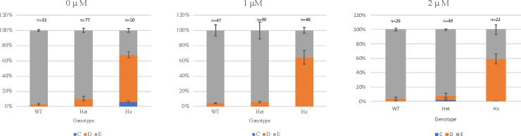
IMAGE

Fig 3

ID
ZDB-IMAGE-190723-1339
Source
Figures for Krall et al., 2019
Image
Figure Caption

Fig 3 Results of phenotypic and genotypic analysis following treatment of in-crossed heterozygous <italic>bmp4</italic><sup><italic>st72/+</italic></sup> zebrafish with 0 μM, 1 μM and 2 μM PTC124.

Larvae were not dechorionated and PTC124 treatment was started at 0 hours post fertilization (hpf). Analysis was performed at 72 hpf and the results are shown as the mean of 3 independent experiments, except for treatment with 2 μM PTC124, which is shown as the mean of 2 independent experiments. Larvae were scored as wildtype (E), mild reduction/deformity in ventral tail fin (D+ or D) or severe reduction/deformity in ventral tail fin (C). WT = wildtype, het = heterozygote, HZ = homozygote. Error bars show standard error of the mean. We tested for trend in the probability of tail fin defects across the increasing doses of 0 μM, 1.0 μM and 2.0 μM PTC124 treated, groups of homozygous larvae [24]. The probability of tail fin defect decreased from 68% to 64.58% and 59.09% as the dose increased to 1.0 μM and 2.0 μM. However, the trend failed to meet the statistical significance (p = 0.4681).

Figure Data
Acknowledgments
This image is the copyrighted work of the attributed author or publisher, and ZFIN has permission only to display this image to its users. Additional permissions should be obtained from the applicable author or publisher of the image. Full text @ PLoS One