IMAGE

Figure 3

ID
ZDB-IMAGE-190723-1212
Source
Figures for Jagau et al., 2019
Image
Figure Caption

Figure 3

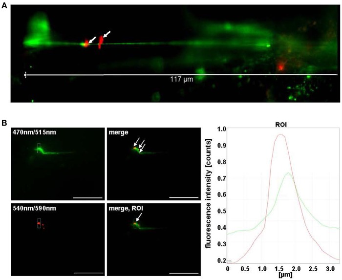
Pneumococci bind to VWF strings generated in continuous flow. (A) HMW VWF fiber generation was microscopically quantified after exposing confluently grown HUVEC to shear stress using a microfluidic device (ibidi®) at 10 dyne/cm2. FITC-conjugated VWF-specific antibodies detected HMW VWF strings. White arrows point to red fluorescent pneumococci attached to long VWF strings. (B) Attachment of RFP-expressing pneumococci to green fluorescent VWF strings was microscopically observed after 30 min in constant flow (white arrows) and was confirmed by software-based evaluation of fluorescence intensities of the defined ROI. Pictures were taken in real time using the fluorescence equipment of a confocal laser scanning microscope (SP8, Leica). Scale bar represents 10 μm.

Acknowledgments
This image is the copyrighted work of the attributed author or publisher, and ZFIN has permission only to display this image to its users. Additional permissions should be obtained from the applicable author or publisher of the image. Full text @ Front Microbiol