IMAGE

Fig. 4

ID
ZDB-IMAGE-190723-109
Source
Figures for Zhang et al., 2019
Image
Figure Caption

Fig. 4

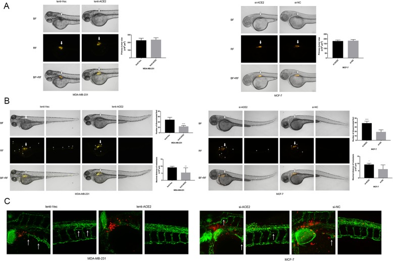
ACE2 inhibits breast cancer angiogenesis in vivo. (a) Equal number of Dil-labelled transfected MDA-MB-231 and MCF-7 cells were microinjected into the perivitelline space of embryos at 48 h post-fertilization. Thick arrows point to the injection sites. BF: bright field; RF: rhodamine fluorescence. (b) Dissemination and metastasis of the tumour cells in the zebrafish at 24 h post-injection (observed under a fluorescence microscope) and quantification of the number of disseminated foci and maximal metastatic distance. Small arrowheads point to the disseminated and metastatic tumour foci (MDA-MB-231, n = 9; MCF-7, n = 6). (c) Vasculature and neo-angiogenesis surrounding the primary tumour and metastasis sites in the zebrafish at 24 h post-injection (observed under a confocal microscope). Thin arrows point to newly formed tumour vessels. Fluorescence microscope magnification: 50×. Confocal microscope magnification: 200×. *P < 0.05; **P < 0.01; ***P < 0.001; ****P < 0.0001

Acknowledgments
This image is the copyrighted work of the attributed author or publisher, and ZFIN has permission only to display this image to its users. Additional permissions should be obtained from the applicable author or publisher of the image. Full text @ J. Exp. Clin. Cancer Res.