
|
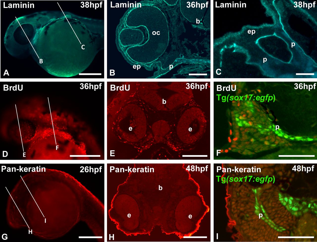
Fig. 3
Whole-mount immunostaining. (A,D,G) Whole-mount, (B,E,H) low- and (C,F,I) high-magnification cross sections. Lines in A,D,G indicate approximate level of sectioning in B,E,H and C,F,I. (A–C) Wild-type (WT) zebrafish immunostained for laminin. Note the basal lamina delimiting optic cup, brain, epidermis and endodermal pouches. (D–F) Tg(sox17:egfp) zebrafish pulse-labeled with BrdU and whole-mount stained with an anti-BrdU antibody. (G-I) Tg(sox17:egfp) zebrafish immunostained with a pan-cytokeratin antibody. The whole-mount images in D,G show the red channel only; the GFP fluorescence is likewise visible prior to dehydration and embedding. b, brain; e, eye; ep, epidermis; oc, optic cup; p, endodermal pouch. Scale bars: (A,D,G) 250 µm; (E,H) 100 µm; (B,F,I) 50 µm; (C) 25 µm.