
Image
|
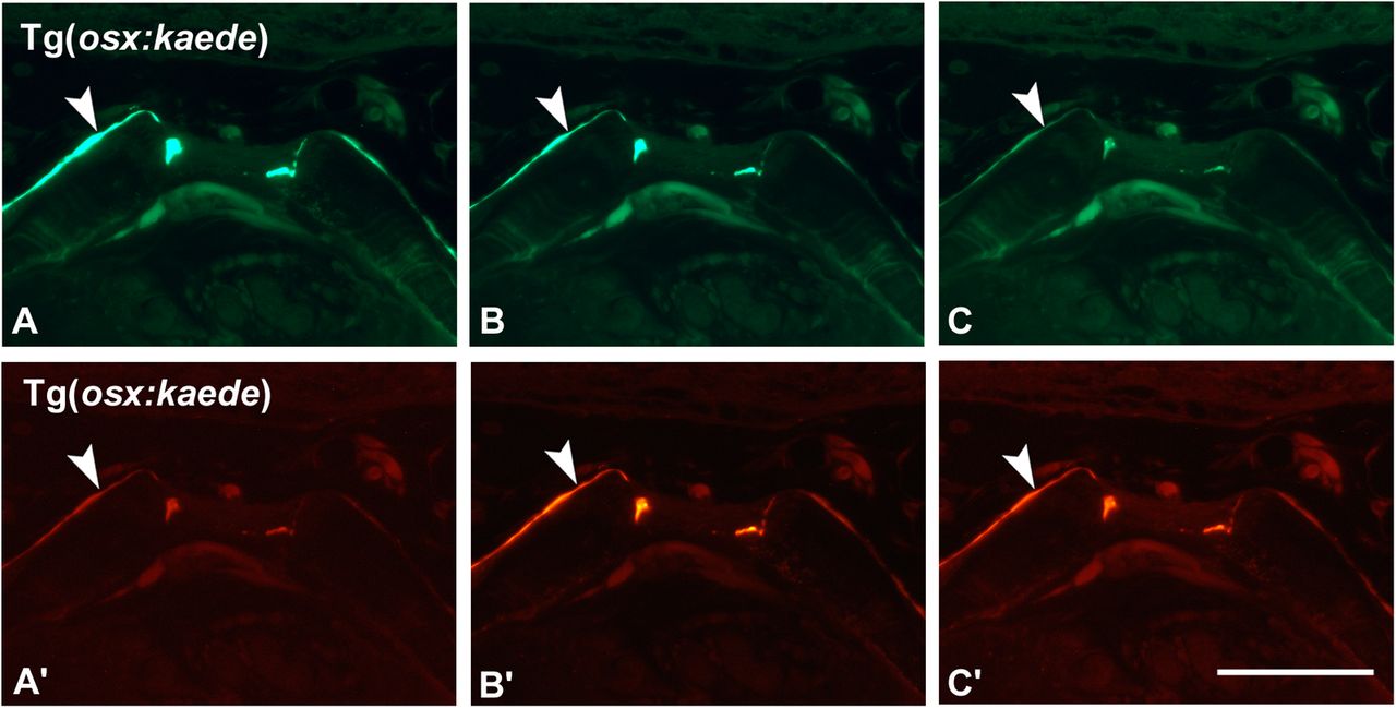
Figure Caption
Fig. 2
Imaging of photoactivatable kaede. Vertebral end plates of an adult Tg(osx:kaede) zebrafish shown using a GFP (A–C) and rhodamine (A′–C′) filter, before (A,A′), after 5 s (B,B′) and 30 s (C,C′) of exposure to a wavelength of 365 nm. Note the fading of the green signal from A to C and strengthening of the red signal from A′ to C′ in the osteoblasts lining the vertebral end plates (arrowheads). All pictures were taken using the same exposure time (1.6 s). Scale bar: 25 µm.
Acknowledgments
This image is the copyrighted work of the attributed author or publisher, and
ZFIN has permission only to display this image to its users.
Additional permissions should be obtained from the applicable author or publisher of the image.
Full text @ Biol. Open