
|
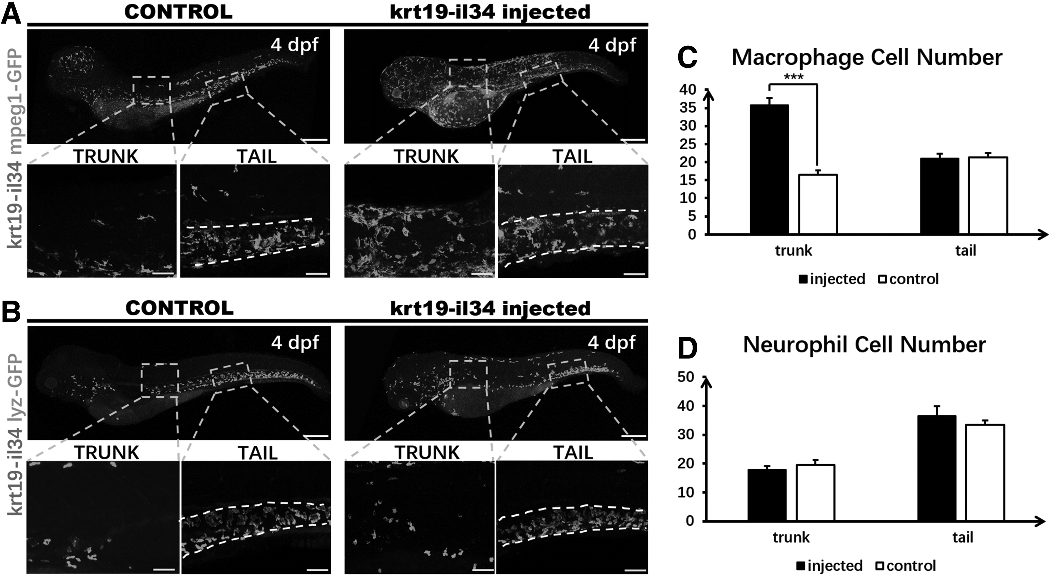
Fig. 1
Transiently overexpressed IL-34 in epidermis led to macrophage enrichment in epidermis. A total of 1.8 nL (30 ng/μL) of the pBLK-krt19-il34-sv40 construct was microinjected into one-cell stage Tg (mpeg1: GFP) (A) and Tg (lyz: EGFP) (B) zebrafish embryos. (A, B) WISH of il34 expression and whole-mount antibody staining of GFP expression in 4 dpf embryo (6 × ). The whole body picture of the fish is made up of two separate images taken by confocal and stitched together in the Photoshop. Insets are high magnification (20 × ) of the corresponding boxed regions (gray dotted rectangles). (C, D) Quantitative analysis of macrophage or neutrophil cell numbers in uninjected and construct-injected embryos' trunk (approximately between the 3rd and 6th somite, shown in gray dotted rectangle), skin, and CHT tail region (approximately between the 13th and 17th somite, shown between two white dotted lines). Data were analyzed by Mann–Whitney U test, ***p < 0.001 compared to control. n = 5, 5 for the 4 dpf injected and control fish. Bars: 200 μm (the line on 6 times objective pictures); 50 μm (the line on 20 times objective pictures). CHT, caudal hematopoietic tissue; dpf, days postfertilization; IL-34, interleukin-34; WISH, whole mount in situhybridization.