
Image
|
Figure Caption
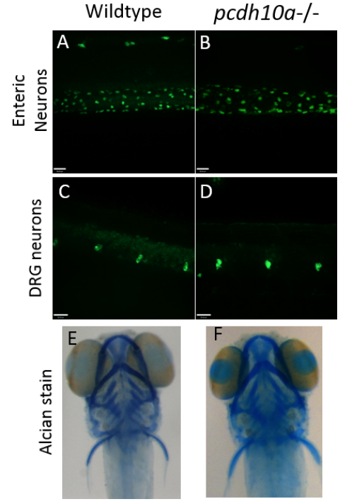
Fig. S5
Removal of pcdh10a does not cause defects in other neural crest derivatives. (A-D) Whole-mount Immunofluorescence with HuC antibody at 5 dpf (A, B) No difference was observed in Enteric neurons between wildtype (A) and pcdh10a -/- (B) embryos. (C,D) No observed difference in Dorsal root ganglia (DRG) between wildtype (C) and pcdh10a -/- (D) embryos. (E, F) Alcian stain at 5 dpf. No observed difference in craniofacial structure between wildtype (E) and pcdh10a -/- (F) embryos.
Acknowledgments
This image is the copyrighted work of the attributed author or publisher, and
ZFIN has permission only to display this image to its users.
Additional permissions should be obtained from the applicable author or publisher of the image.
Reprinted from Developmental Biology, 444 Suppl 1, Williams, J.S., Hsu, J.Y., Rossi, C.C., Artinger, K.B., Requirement of zebrafish pcdh10a and pcdh10b in melanocyte precursor migration, S274-S286, Copyright (2018) with permission from Elsevier. Full text @ Dev. Biol.