
Image
|
Figure Caption
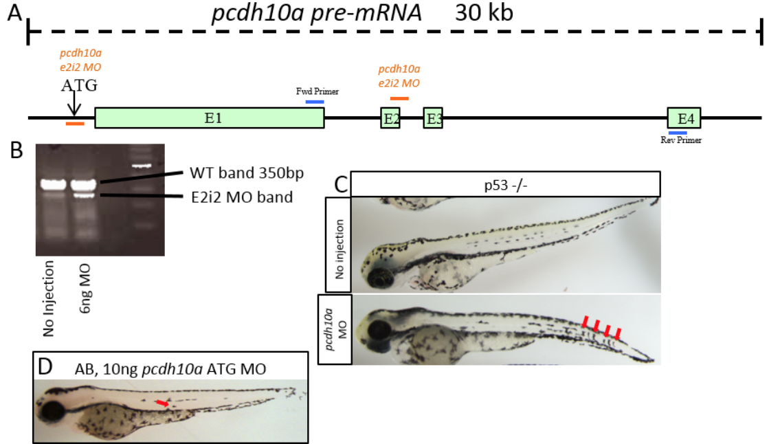
Fig. S1
Pcdh10a Morpholino causes deletion of exon 2. (A) Schematic of pcdh10a pre-mRNA with ATG and e2i2 MO site and PCR primer binding sites. (B) RT-PCR to confirm spice site change in e2i2 MO-injected embryos, in this case, a deletion of exon 2. (C) 6ng E2i2 MO injection into p53 mutant fish shows the same phenotype with internal pigment cells (red arrows). (D) 10ng pcdh10a ATG Morpholino injected into AB mildly phenocopies E2i2 MO with internal pigment cells (red arrow).
Figure Data
Acknowledgments
This image is the copyrighted work of the attributed author or publisher, and
ZFIN has permission only to display this image to its users.
Additional permissions should be obtained from the applicable author or publisher of the image.
Reprinted from Developmental Biology, 444 Suppl 1, Williams, J.S., Hsu, J.Y., Rossi, C.C., Artinger, K.B., Requirement of zebrafish pcdh10a and pcdh10b in melanocyte precursor migration, S274-S286, Copyright (2018) with permission from Elsevier. Full text @ Dev. Biol.