
|
Fig. 1
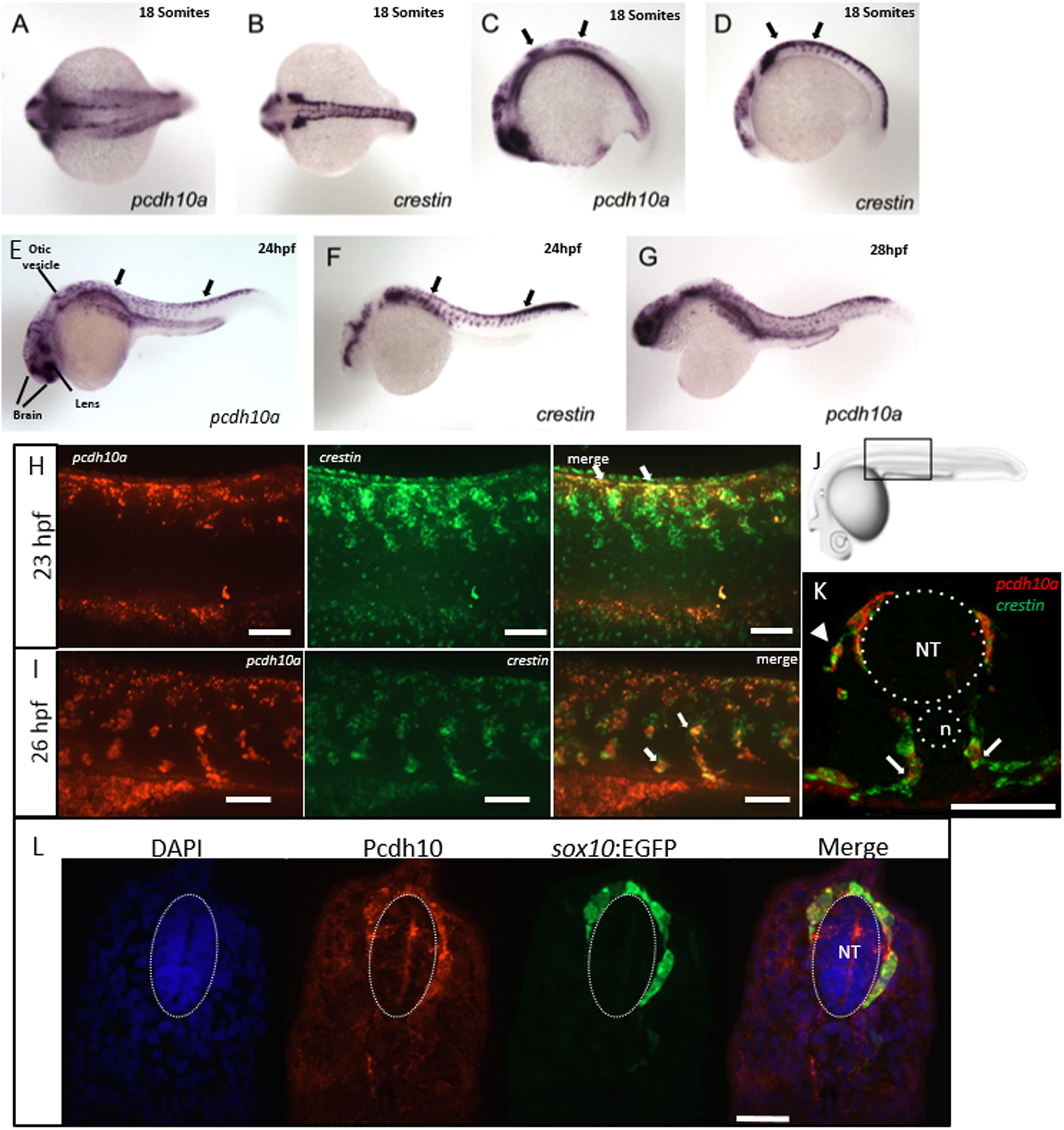
pcdh10a is expressed in a subset of crestin positive migratory neural crest cells (NCCs). (A-G) Whole-mount in situ hybridization (ISH) of pcdh10a and crestin expression during NCC migration in wildtype zebrafish embryos. (A) pcdh10a is expressed in a similar pattern to the NCC marker crestin (B) Dorsal views at 18 somites. (C,D) Lateral views at 18 somites. (E, F) Lateral views at 24 hpf. (G) Lateral view at 28 hpf. (H, I, K) Confocal micrograph Z-stack projections of double Fluorescent ISH (FISH) at 23hpf and 26 hpf respectively in wildtype zebrafish embryos coexpressing pcdh10a (Red) and crestin (Green). Lateral views with dorsal top, ventral down. White arrows mark examples of colocalization. Scale bars are 50 µm. (J) Area of imaging for H, I, K, L. (K) Representative image of a 12 µm cross-section of the embryo in (I). White arrows point to areas of colocalization, and white arrowheads point to NCCs in the dorsolateral pathway. (L) 12 µm cross-section through the trunk of an embryo stained for Pcdh10 (Red) immunofluorescence, with neural crest cells expressing sox10:EGFP (Green). Scale bar is 20 µm, images are a single 1 µm Z-plane. NT; Neural Tube, n; Notochord. Anterior is to the left in all whole mount images.
Reprinted from Developmental Biology, 444 Suppl 1, Williams, J.S., Hsu, J.Y., Rossi, C.C., Artinger, K.B., Requirement of zebrafish pcdh10a and pcdh10b in melanocyte precursor migration, S274-S286, Copyright (2018) with permission from Elsevier. Full text @ Dev. Biol.