
|
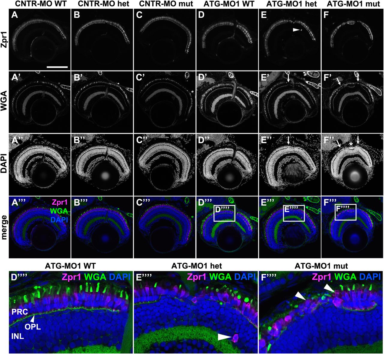
Fig. 5
Knockdown of Lgl2 in pen/lgl2 clutches leads to disorganization of retinal lamination. Immunostaining of transverse retinal sections at 5 dpf of MO-injected and genotyped pen/lgl2 clutches. (A–C″′) CNTR-MO-injected fish and (D-F′′′′) ATG-MO1-injected fish. Zpr1 (A–F) stains double cone cell bodies and WGA (A′–F′) the OPL and OSs. In ATG-MO1-injected hetero- (E–E′′′′) or homozygous (F–F′′′′) fish, layering in the distal retina is disturbed. Arrows in E′, E″, F′ and F″ mark disorganized clusters separated by normally aligned cells (asterisk). Boxed areas in D′′′–F′′′ show enlarged views of PRCs in MO-injected pen/lgl2 WT (D′′′′), heterozygote (E′′′′) and mutant (F′′′′) fish. Occasionally Zpr1 positive cells were seen outside of the PRC layer (E,E′′′′, arrowheads). Arrowheads in F′′′′ mark apically displaced PRCs that are localized on top of another PRC. PRC, photoreceptor layer; OPL, outer plexiform layer; INL, inner nuclear layer. Scale bar: 100 µm.