
IMAGE
Fig. 3
Image
|
Figure Caption
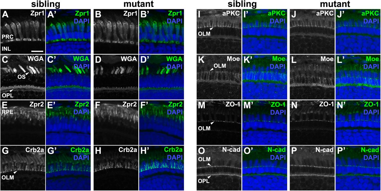
Fig. 3
pen/lgl2 mutant distal retina displays no significant abnormalities in polarity or cellular morphology.Immunostaining of transverse retinal sections at 5 dpf. (A,C,E,G,I,K,M,O) Sibling and (B,D,F,H,J,L,N,P) mutant fish. (A–B′) Zpr1, (C–D′) WGA, (E–F′) Zpr2, (G–H′) Crb2a, (I–J′) aPKC, (K–L′) Moe, (M–N′) ZO-1 and (O–P′) N-cadherin immunostaining of distal retina. PRC, photoreceptor layer; INL, inner nuclear layer; OS, outer segment; OPL, outer plexiform layer; RPE, retinal pigment layer; OLM, outer limiting membrane. Scale bar: 10 µm.
Figure Data
Acknowledgments
This image is the copyrighted work of the attributed author or publisher, and
ZFIN has permission only to display this image to its users.
Additional permissions should be obtained from the applicable author or publisher of the image.
Full text @ Biol. Open