
|
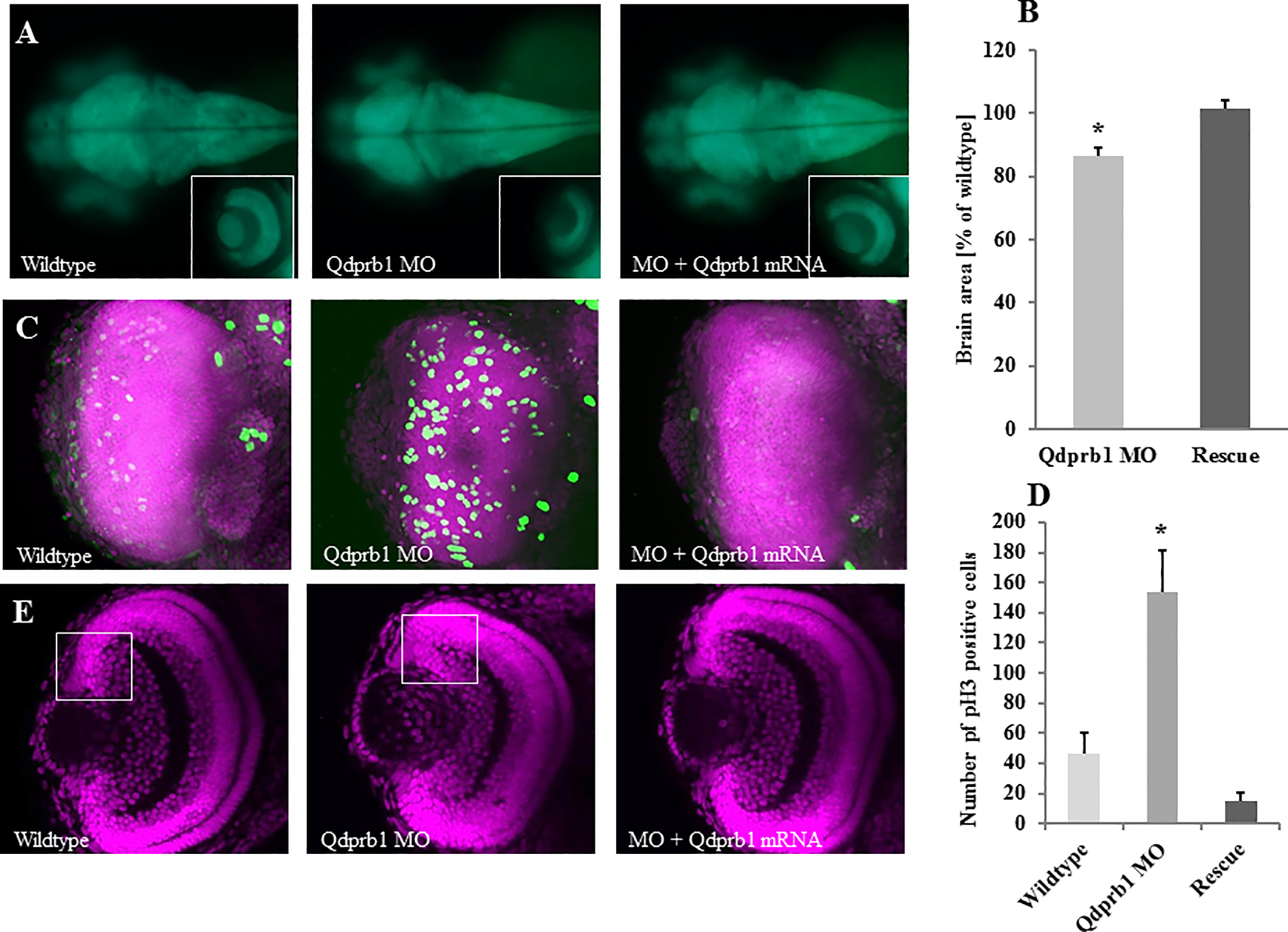
Fig. 4
(A) Dorsal views, anterior to the left. At 3 dpf tg(HuC/D:GFP) transgenic zebrafish show a decreased size of the optic tectum and eye upon qdprb1 knockdown, which can be rescued by co-injecting qdprb1 mRNA. The insets show dorsal views of the left eye, which is reduced in size but still layered upon qdprb1 suppression. This phenotype is rescued upon co-injection of qdprb1 mRNA. The overall GFP signal remains unchanged. (B) The brain area of Qdprb1 hypormorphic embryos is reduced by about 15% compared to wildtypes and rescued by co-injection of qdprb1 mRNA. (C) Z-stack overlays of DAPI (pink) and pH3 (green) staining of dorsally imaged retinas reveal an increased number of pH3-positive retinal cells in Qdprb1 hypormorphic embryos (3 dpf), which are not restricted to the CMZ like found in wildtype retinas. Number and distribution of pH3-positive cells is restored by co-injection of qdprb1 mRNA. (D) Statistical analysis shows a significant increase in pH3 positive cells upon qdprb1knockdown (n = 6) in comparison to wildtype zebrafish (n = 7), which could be rescued by addition of qdprb1 mRNA (n = 3). (E) Z-confocal image of DAPI staining (pink) of the retina at 3 dpf shows retinal layers although decreased overall size and broadened CMZ in qdprb1 hypomorphic embryos.