
|
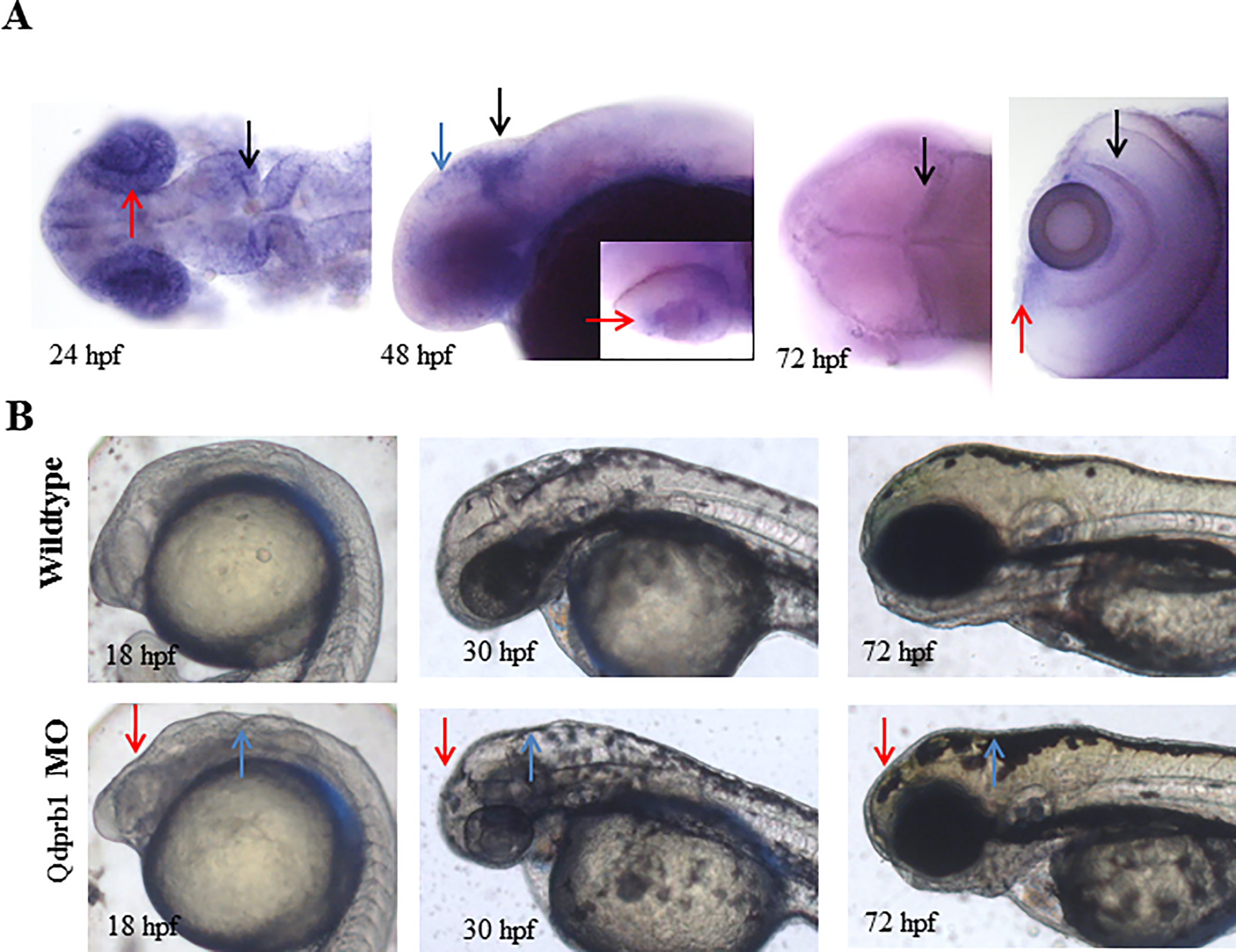
Fig. 2
(A) WISH of qdprb1 at 24 hpf (dorsal view, anterior to the left) shows staining in eye (red arrow) and mid-hindbrain boundary (black arrow), at 48 hpf (lateral view, anterior to the left) in the optic tectum (blue arrow) and the mid-hindbrain boundary (black arrow) and CMZ (inset; red arrow, dorsal view, anterior to the left), and at 72 hpf (dorsal view, anterior to the left) in proliferative regions of the optic tectum (black arrow, left picture), CMZ (red arrow, right picture, dorsal view of the eye), as well as inner retinal layer (black arrow). (B) Lateral views, anterior to the left. Comparison of wildtype embryos at 18 hpf, 24 hpf and 72 hpf and qdprb1 hypomorphic embryos exhibit abnormal midbrain (red arrow) and anterior hindbrain (blue arrow) morphology and microcephaly.