
|
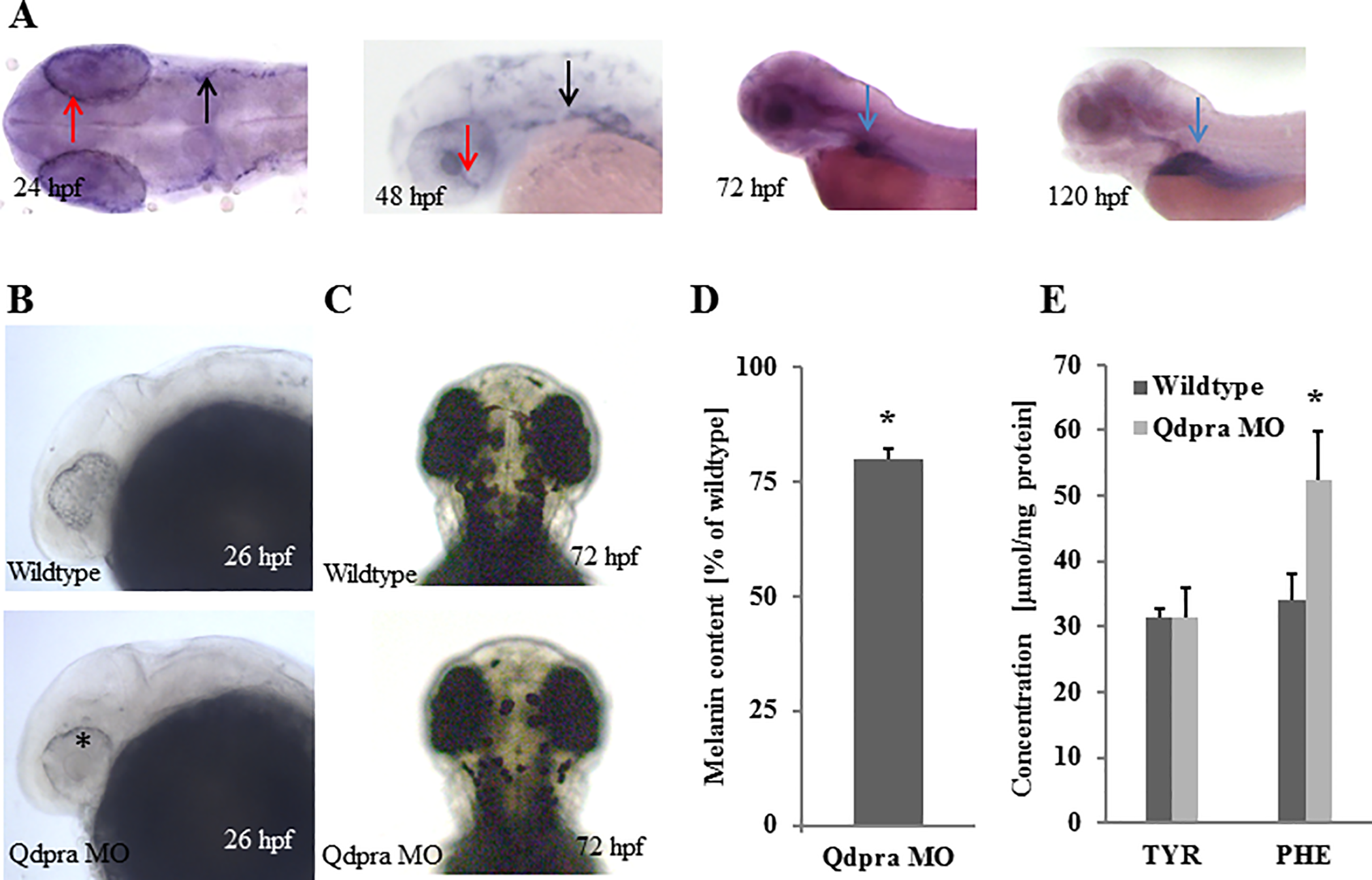
Fig. 1
(A) WISH of qdpra at 24 hpf (dorsal view, anterior to the left) shows staining in retinal pigment epithelium (red arrow) and neural crest cells/melanophore precursor (black arrow). At 48 hpf qdpra transcripts are found in the retinal pigment epithelium, choroid fissure (red arrow) and neural crest cells (black arrow). At 72 hpf and more pronounced at 120 hpf (lateral views, anterior to the left), staining is present in the liver (blue arrow). (B) Lateral views with anterior to the left and (C) dorsal views with anterior to the top of embryos at stages indicated. Knockdown of qdpra results in reduced pigments in the eye (asterisk) at 26 hpf (B) and overall diminished pigmentation at 72 hpf (C). (D) At 72 hpf melanin content is reduced by 20% (of wildtype) in Qdpra hypomorphic zebrafish. (E) Amino acid analysis shows hyperphenylalaninemia and normal tyrosine upon qdpraknockdown.