
|
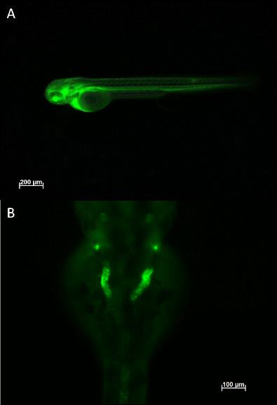
Fig. 3
Injection of 10 kDa FITC-dextran into a wild-type zebrafish embryo at 72 h post-fertilisation visualised on a fluorescence inverted compound microscope. a Injection of the tracer into the common cardinal veins taken just after injection, showing uptake of the dextran into the blood vasculature of the embryo. b Dorsal visualisation of the dextran being taken up in to the pronephric duct and tubules after 20 h incubation post injection. Note that the tubules appear convoluted as they form a coiled loop around the glomerulus (not shown) and show reduced fluorescence in the rest of the embryo indicative of normal renal filtration processes