
|
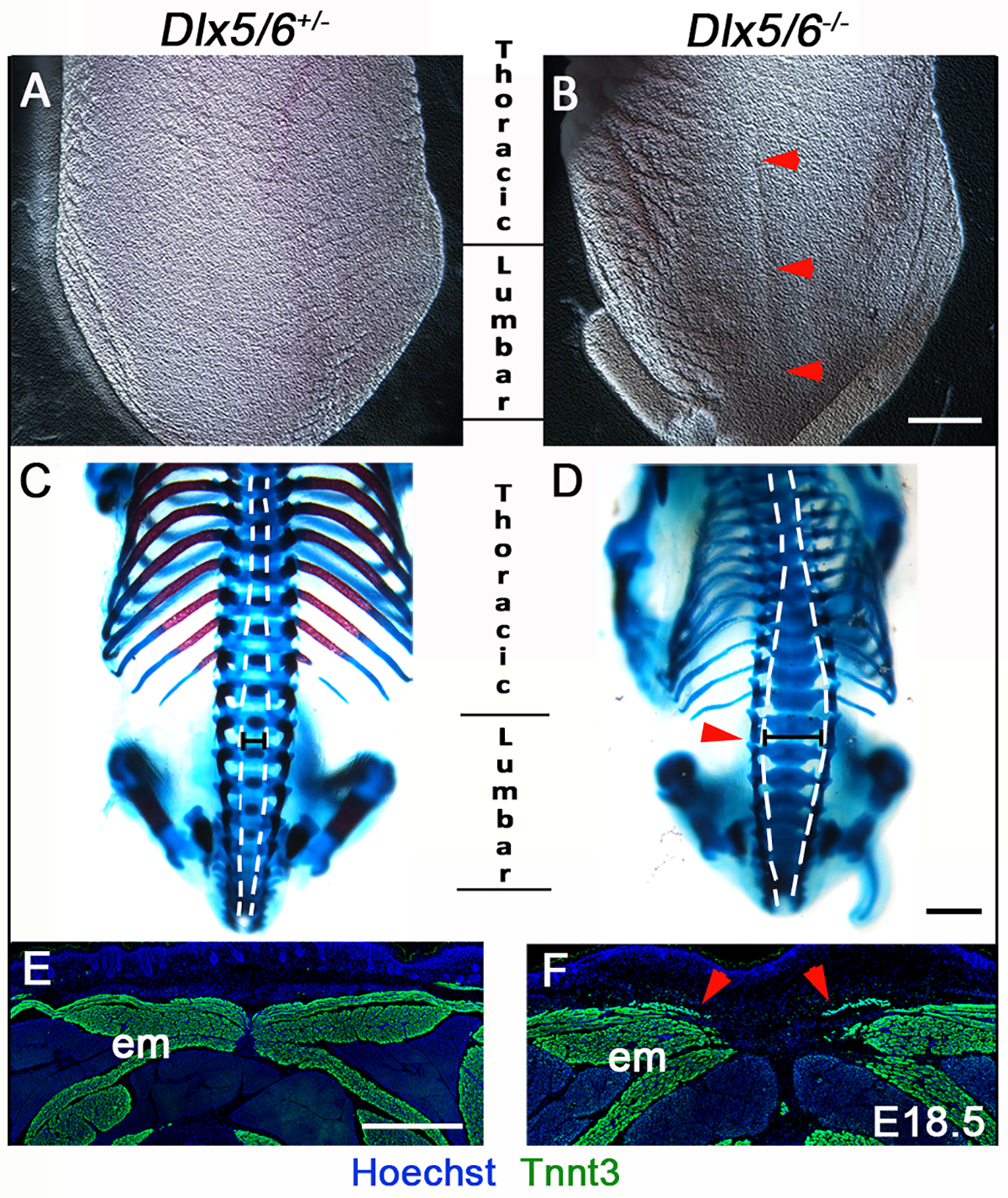
Fig. 2
Dorsal midline defects in perinatal Dlx5/6-/- mice.
(A-D) Macroscopic dorsal view (A-B) and skeletal preparation (C-D) of the posterior axis of control Dlx5/6+/- and Dlx5/6-/- mutant mice at E18.5 (n = 6 for each condition). (E-F) Immunostaining on coronal cryosections for Tnnt3 in dorsal musculature of control Dlx5/6+/- and Dlx5/6-/- E18.5 foetuses (n = 3 for each condition). Dlx5/6-/- mutants display a dorsal split already evident at macroscopic inspection (B, red arrowheads). This phenotype is associated with defects of thoracic/lumbar vertebrae (D, red arrowhead) and of epaxial muscle formation at the dorsal midline (F, red arrowheads). Abbreviations: em, epaxial muscles. Scale bars in B and D 2000 μm and in E 200 μm