
Image
|
Figure Caption
Fig. S1
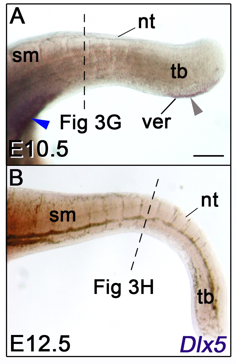
(A, B) Lateral views of whole-mount in situ hybridization for Dlx5 in E10.5 and E12.5 mice. The dashed lines indicate the section levels analysed in Fig 3G and 3H. Dlx5 expression is detected at the cloacal level and in the VER at E10.5 (A, blue and grey arrowheads respectively) but is not detectable at E12.5. Abbreviations: nt, neural tube; sm, somitic mesoderm; tb, tail bud; ver, ventral ectodermal ridge. Scale bar in A for A-B 200 μm.
Acknowledgments
This image is the copyrighted work of the attributed author or publisher, and
ZFIN has permission only to display this image to its users.
Additional permissions should be obtained from the applicable author or publisher of the image.
Full text @ PLoS One