
|
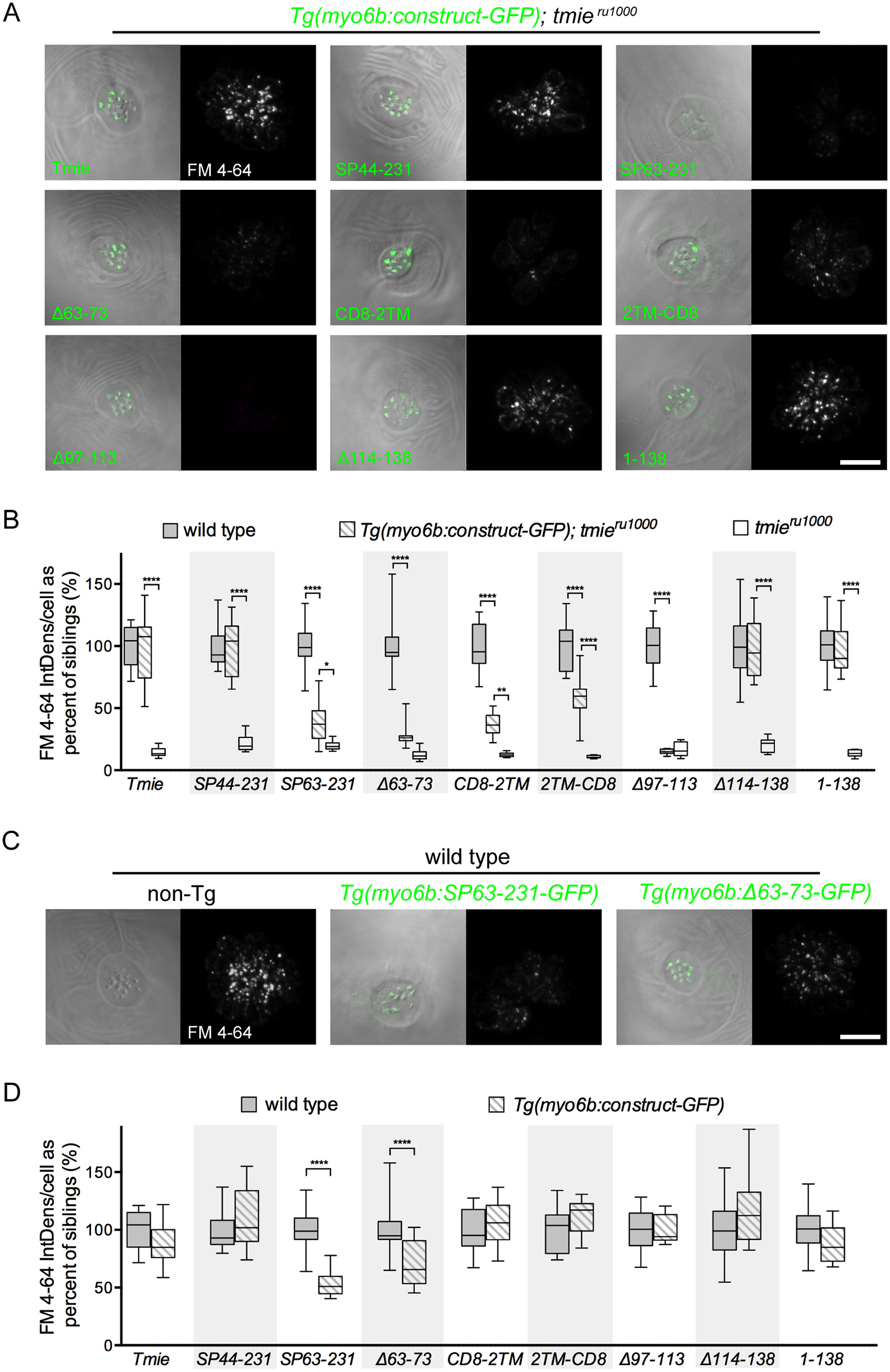
Fig. 6
All images are a top-down view of a representative neuromast from 6 dpf larvae collected using confocal microscopy. The left image is a single plane through the stereocilia (green dashed line in Fig 1G) with DIC + GFP fluorescence. The right image is a maximum projection of the 7 sections in the soma region (magenta bracket in Fig 1G) showing FM 4–64 fluorescence. (A) Representative images of neuromasts in tmieru1000 larvae, each stably expressing an individual tmie construct. FM fluorescence was normalized to wild type non-transgenic larvae generated with the Tmie-GFP line. (B) Box-and-whiskers plot of the integrated density of FM fluorescence/cell for each tmieconstruct. We normalized values to the average of wild type siblings for each construct. (C) Representative images of neuromasts in wild type larvae with or without transgene. FM fluorescence was normalized to wild type non-transgenic larvae of the Tmie-GFP line. (D) Box-and-whiskers plot of the integrated density of FM fluorescence/cell in wild type neuromasts with and without tmie transgene. We normalized values to the average of wild type siblings for each construct. Significance determined within each clutch by one-way ANOVA, n ≥ 9, **p < 0.01, ***p < 0.001, ****p < 0.0001. Scale bars are 10μm.