
Image
|
Figure Caption
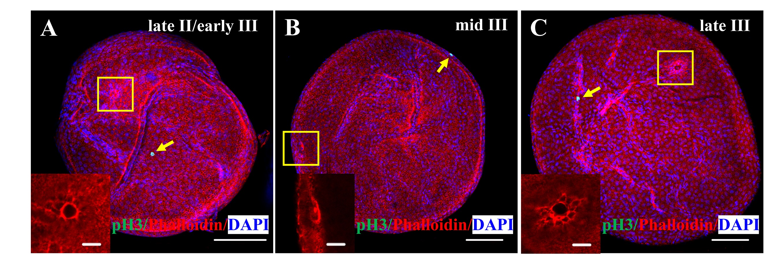
Fig. S3
The micropylar cell is pH3 negative.
Immunofluorescence of F-actin and pH3 in wild type oocytes. The pH3 signal was not found in the micropylar cell (n = 70 oocytes). Insets are high magnification images of the micropyle in the yellow boxed area. Yellow arrow, pH3 positive cell, Scale bar, 100 μm; insets, 20 μm.
Acknowledgments
This image is the copyrighted work of the attributed author or publisher, and
ZFIN has permission only to display this image to its users.
Additional permissions should be obtained from the applicable author or publisher of the image.
Full text @ PLoS Genet.