
Image
|
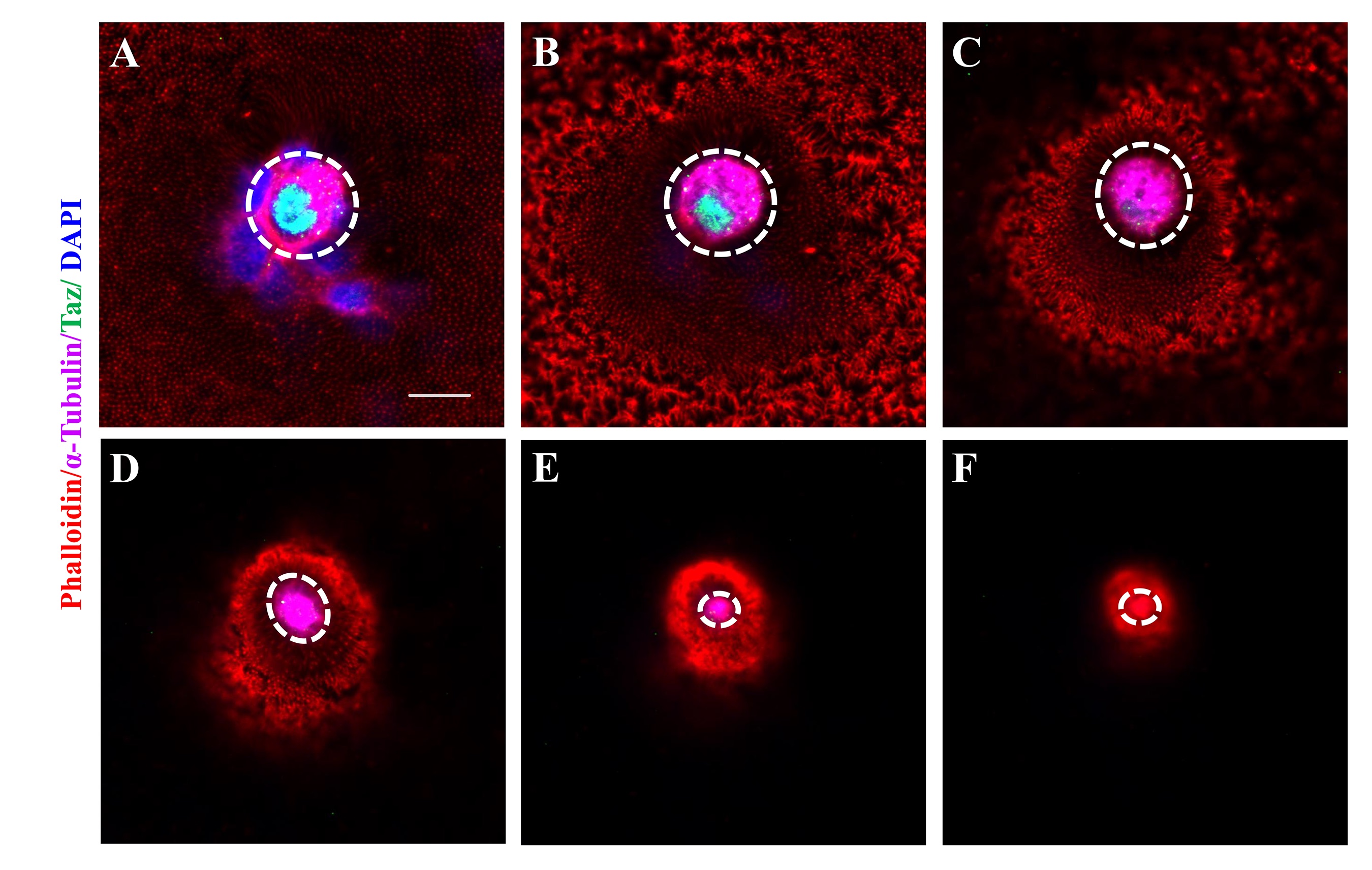
Figure Caption
Fig. S2
Cytoskeleton in the micropylar cell.
Consecutive confocal sections at 2.5 μm intervals showing immunofluorescence of Taz, F-actin and α-Tubulin in a stage III wild type oocyte (n = 17). (A-B) show the micropylar cell body in which Taz is mainly expressed in the nucleus and α-Tubulin is in the cytoplasm. (C-F) show the cytoplasmic extension of the micropylar cell; α-Tubulin is enriched in the cytoplasm and F-actin is deposited at the leading tip (F). Dashed white circle, the micropylar cell. Scale bar, 10 μm.
Acknowledgments
This image is the copyrighted work of the attributed author or publisher, and
ZFIN has permission only to display this image to its users.
Additional permissions should be obtained from the applicable author or publisher of the image.
Full text @ PLoS Genet.