
|
Fig. S1
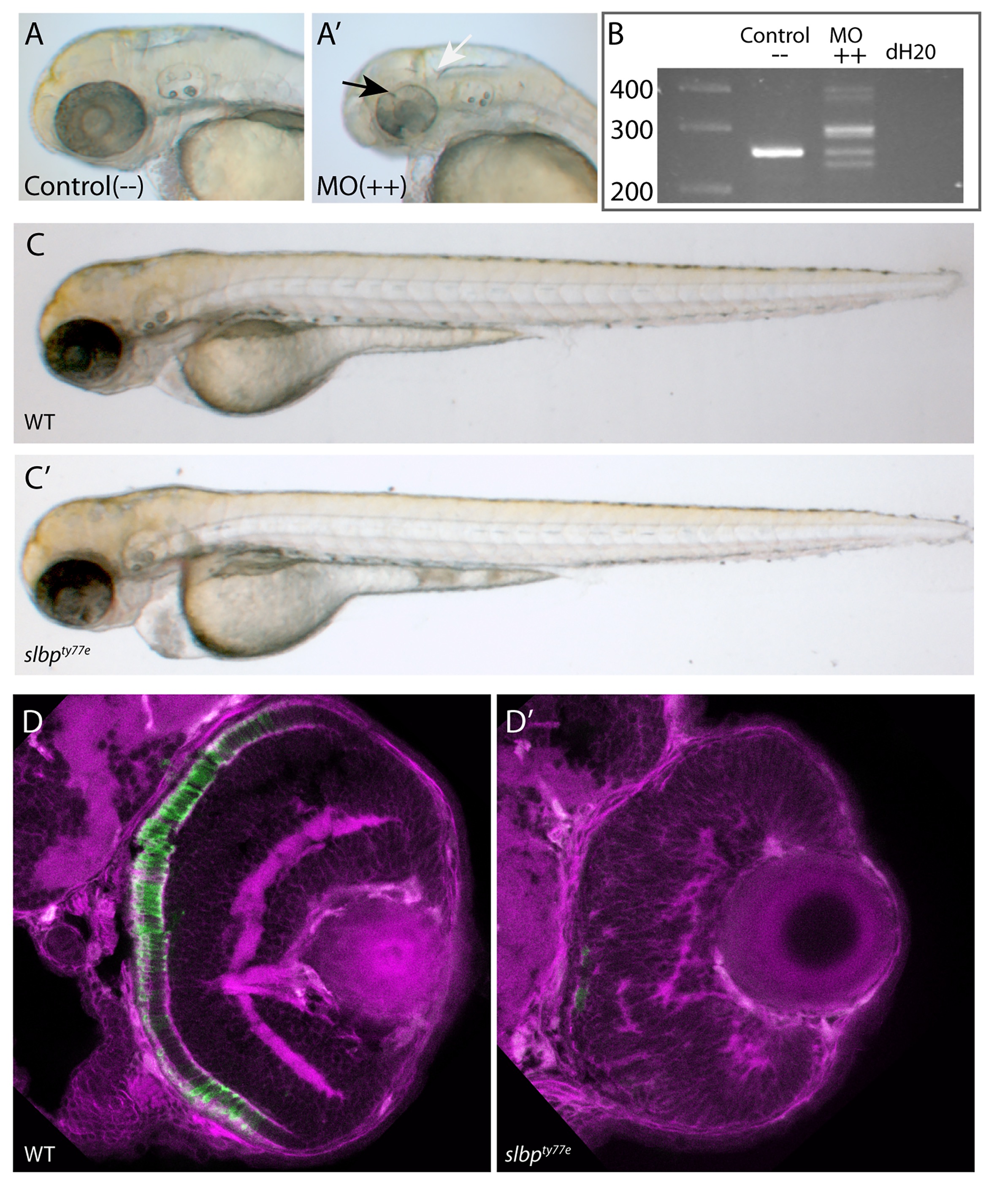
(A-A’) Analysis of live 50hpf Control (A) and Morpholino injected embryos (A’) show that the morphological dent caudal to the MHB (white arrow), indentations in the retina (black arrow) and coloboma are successfully phenocopied.
(B) Electrophoresis gel of RT-PCR analysis confirmed that several missplicing events occur as at least five variably sized products were generated (++ lane).
(C-C’) Injection of degradation-resistant slbpTT-AA-RFP synthetic RNA into WT(C) and ele(C’)mutants rescues ele phenotype. Note heart oedema still present in some cases (C’).
(D-D’) Frontal sections of 3dpf wildtype (D) and slbpty77e (D’) retinas showing anti-γ-tubulin labelled neurites/neuropil (magenta) and Rho4D2-expressing rod photoreceptors (green).
(TIF)