
|
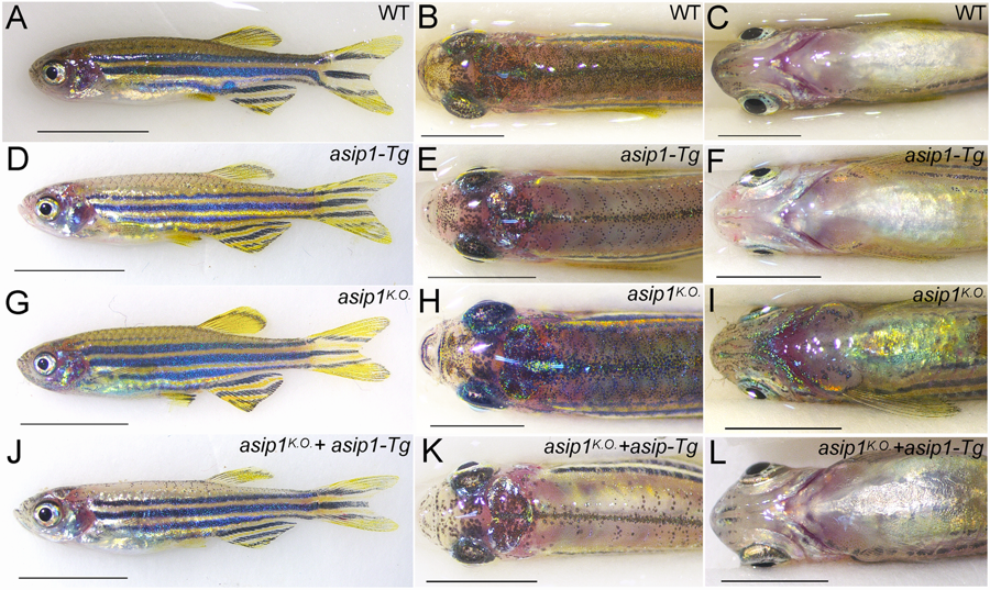
Fig. 7
Functional rescue of CRISPR-mediated asip1 mutation. Lateral (A,D,G,J), dorsal (B,E,H,K) and ventral-belly (C,F,I,L) views of 160 dpf WT, asip1-Tg, asip1K.O., and asip1K.O; asip1-Tg zebrafish. The pigment pattern of WT zebrafish shows (A) normal striped pattern, (B) dark dorsum and (C) light belly. The pigment pattern of asip1-Tg fish shows (D) almost normal striped pattern, although dark stripe 2D??? is rather thinner???, (E) hypopigmented dorsum and (F) light belly. The pigment pattern of asip1K.O. fish shows (F) almost normal striped pattern, but with dark stripes 2 V and 3 V more developed than WT fish, (H) pigmented dorsum similar to WT and (I) hyperpigmented belly compared to WT. The asip1K.O + asip1 − Tg phenotype shows a phenotype similar to the asip1 − Tg zebrafish, except that dark stripe 2D is more prominent. Scale bar: 5 mm.