
|
Fig. 4
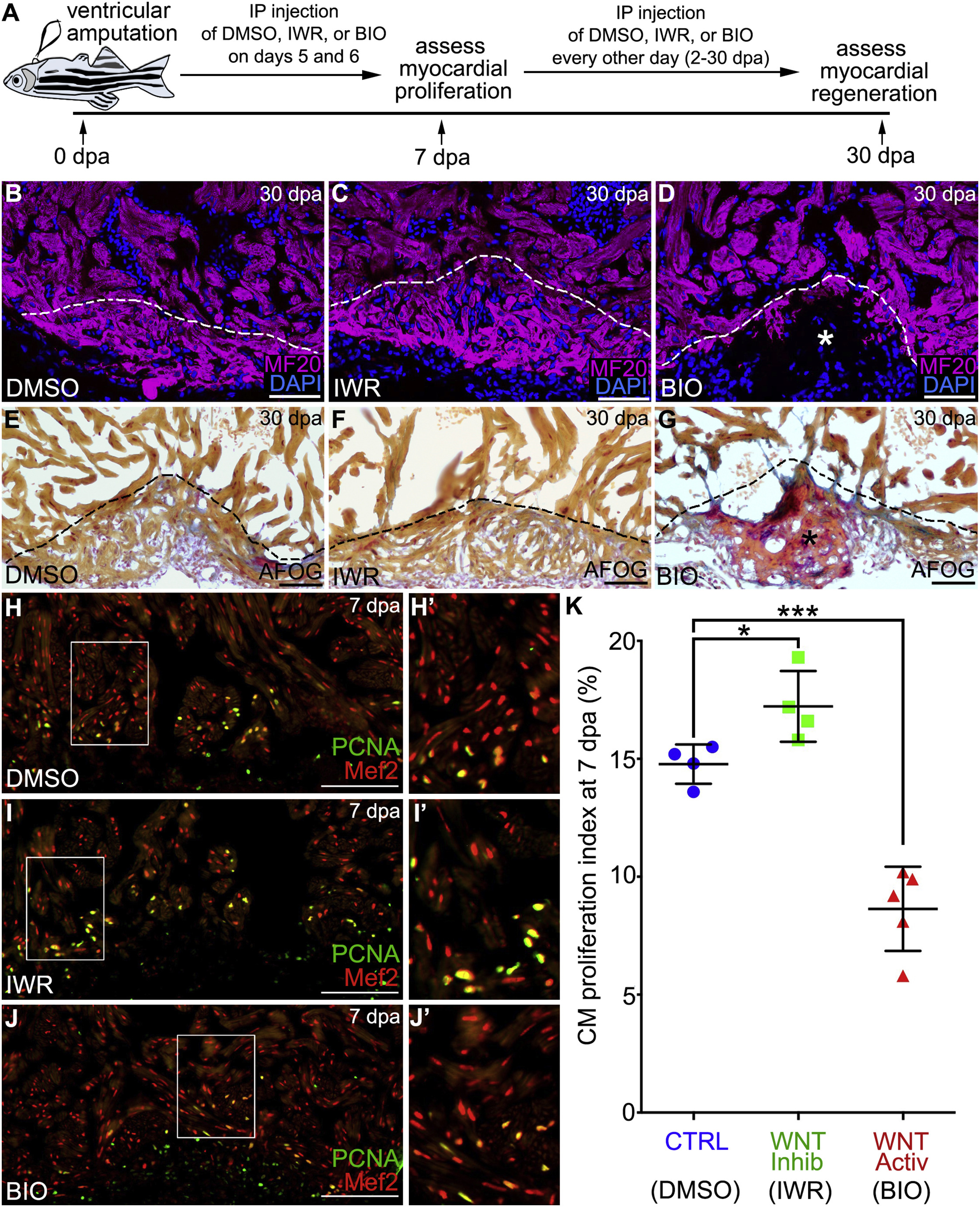
Inhibition of Wnt Signaling Is Required for ZebrafishHeart Regeneration by Bolstering Myocardial Proliferation
(A) Experimental strategy used to inhibit or augment Wnt signaling during the regenerative window. Injured animals were injected intraperitoneally (IP) with DMSO, IWR, or BIO on 5 and 6 dpa before cardiomyocyte proliferation was measured on 7 dpa. Myocardial regeneration was examined on 30 dpa followed by IP injection of DMSO, IWR, or BIO every other day throughout 2–30 dpa.
(B–G) Cardiac sections from 30 dpa wild-type animals injected with DMSO (B and E), IWR (C and F), or BIO (D and G). Single confocal slices of sections immunostained with MF20 (magenta) and counterstained with DAPI (blue) are shown in (B)–(D). Sections stained with AFOG to visualize muscle (brown), fibrin (red), and collagen (blue) are shown in (E)–(G). Asterisks in (D) and (G) highlight gaps in the myocardial wall filled with collagen-rich scar tissue. Heart regeneration occurred in 7/8 DMSO-treated, 4/7 IWR-treated, and 2/8 BIO-treated animals.
(H–J′) Cardiac sections from 7 day post-amputation wild-typeanimals treated with DMSO (H and H′), IWR (I and I′), and BIO (J and J′) were double immunostained to detect cardiomyocyte nuclei (Mef2 antibody; red) and cycling cells (α-PCNA antibody; green). Compound microscopic images are shown.
(K) Bar graph showing cardiomyocyte proliferation indices for the indicated experimental groups (DMSO, n = 4; IWR, n = 4; BIO, n = 5). 4–6 sections per heart were analyzed and averaged to generate each data point. Statistical significance was determined using a Student’s t test. Error bars: ±1 SD. ∗p < 0.05. ∗∗∗p < 0.001. Scale bars: 50 μm.