
|
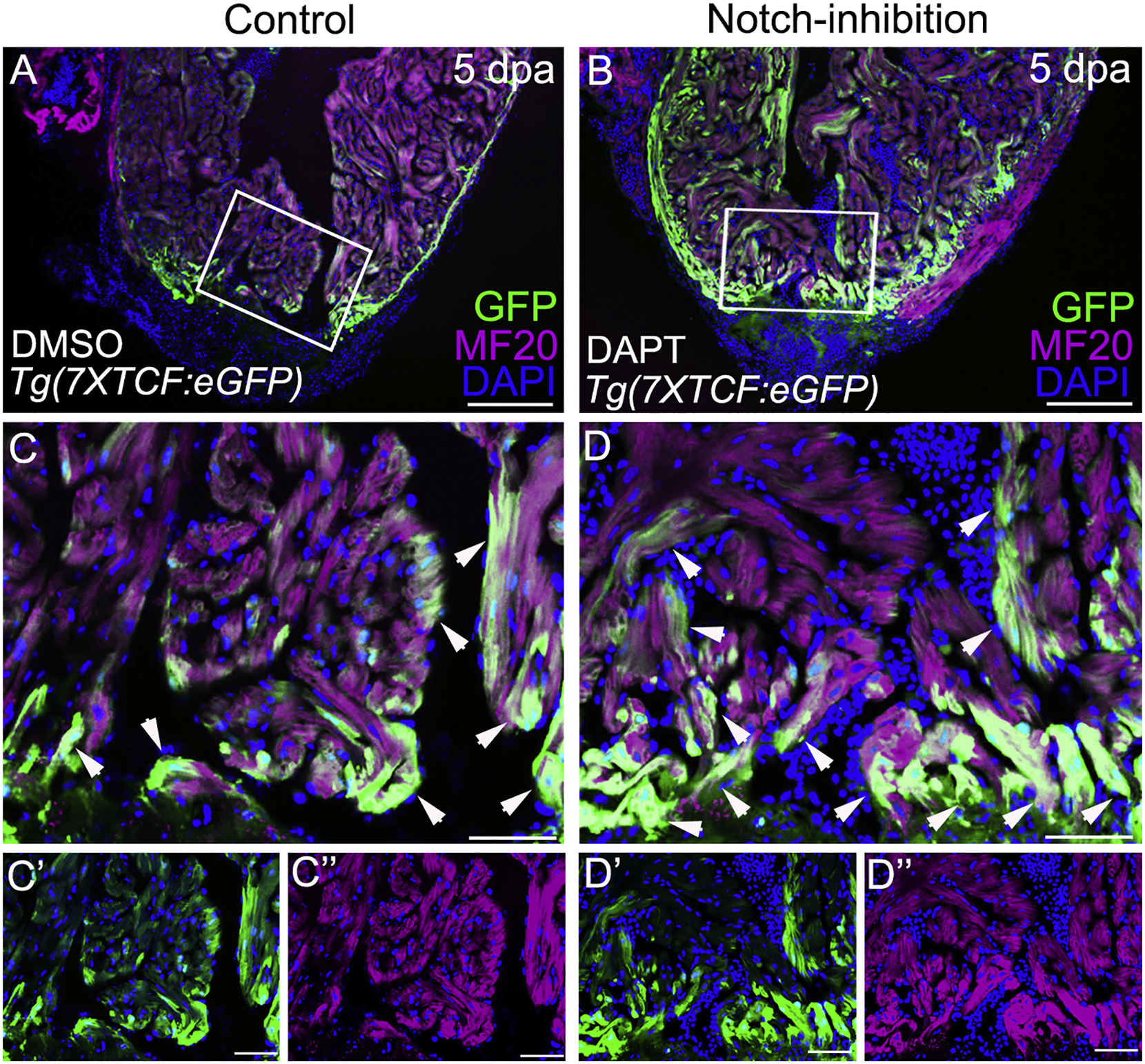
Fig. 3
Notch Inhibition Boosts Wnt Signaling in the Myocardium of Injured Zebrafish Hearts
(A and B) Confocal slices of cardiac sections from 5 dpa Tg(7xTCF:eGFP) animals injected daily with DMSO (A) or N-[N-(3,5-Difluorophenacetyl)-L-alanyl]-S-phenylglycine t-butyl ester (DAPT) (B). Animals were double immunostained to detect GFP (green) and myocardium (MF20 antibody; magenta) and counterstained with DAPI (blue).
(C–D″) Boxed regions in (A) and (B) are shown at higher magnification in (C) and (D). Split channels are shown in (C′), (C″), (D′), and (D″). Arrows point to myocardial cells with active Wnt signaling. n = 3 hearts per experimental group. At least 3 sections were examined per heart. Little to no variability was observed between animals in each experimental group.