
|
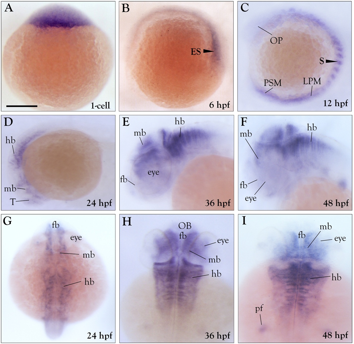
Fig. 1
Expression of zebrafish peli1b is spatiotemporally restricted during embryonic development. (A–C) Lateral view of early stage embryos. (D–F) Lateral view and (G–I) dorsal view of late stage embryos. (A) Animal-pole view of an embryo showing that peli1b transcripts are abundant. (B) Animal-pole view of a shield stage embryo showing a greater level of peli1b transcripts on the dorsal side than on the ventral side. (C) At 12 hpf, there is peli1b expression in the hindbrain, somites, and presomitic mesodermregion. At 24 hpf, the lateral view (D) and dorsal view (G) show abundant peli1b expression in the telencephalon, diencephalon, and hindbrain (r 1-7), but a lack of expression in the midbrain–hindbrain boundary (MHB). At 36 hpf, the lateral view (E) and dorsal view (H) show abundant peli1b transcripts in the diencephalon, hindbrain, eyes, and pectoral fins. At 48 hpf, the lateral view (F) and dorsal view (I) show peli1b expression in the diencephalon, hindbrain (r 1-7), eyes, and pectoral fins. In the colorimetric experiments, samples were incubated for 3 h at room temperature. Abbreviations: ES, embryonic shield; OP, Optic premordium; LPM, lateral plate mesoderm; S, somites; PSM, pre-somitic mesoderm; hb, hindbrain; Mb, midbrain; T, telencephalon; fb, forebrain; OB, olfactory bulb. (n = 3). Scale bar- 50 μm.
Reprinted from Gene, 694, Kumar, A., Anuppalle, M., Maddirevula, S., Huh, T.L., Choe, J., Rhee, M., Peli1b governs the brain patterning via ERK signaling pathways in zebrafish embryos, 1-6, Copyright (2019) with permission from Elsevier. Full text @ Gene