
|
Fig. 6
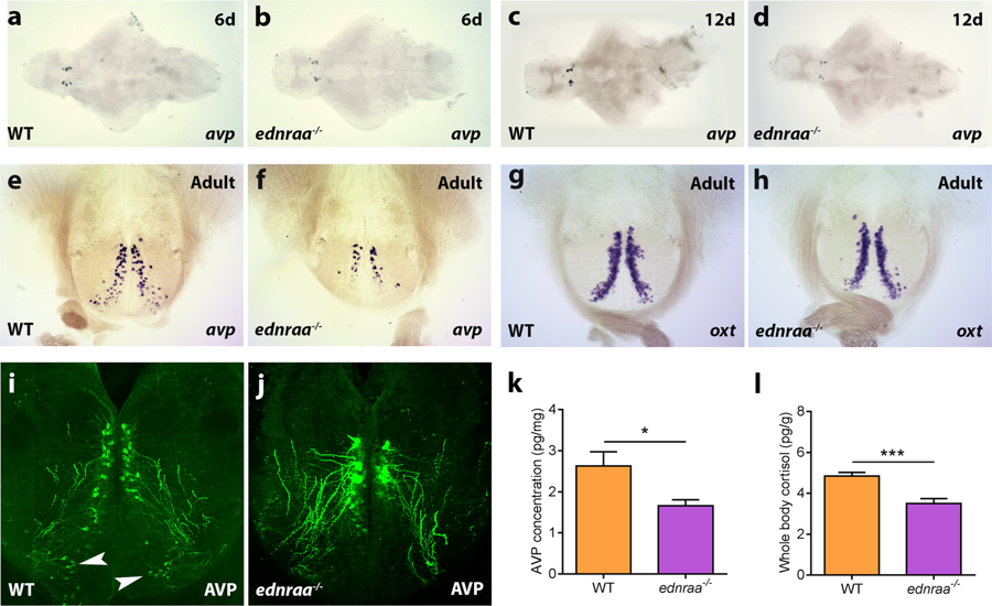
Altered distribution of arginine vasopressin neurons in ednraa−/−. In situ hybridisation showing that expression of arginine vasopressin (avp) is similar in the brain of WT (a,c,e) and ednraa−/− (b,d,f) at 6 days (a,b) and 12 days (c,d). (e,f) Reduced expression of avp in the ventral parvocellular preoptic area of ednraa−/− of adult fish. (g,h) In situ hybridisation showing that oxytocin (oxt) expression is similar in WT and ednraa−/− adult fish. (i,j) Anti-AVP antibody staining shows reduced labelling in the parvocellular preoptic area of ednraa−/− (j) compared to WT (i) (arrowheads). Dorsal magnocellular neurons have a larger cell body and thicker projections. (k) Reduced levels of AVP in the brain of ednraa−/− compared to WT. (l) Decreased whole-body cortisol levels in ednraa−/− compared to WT. *p < 0.05, ***p < 0.001.