
|
Fig. S1
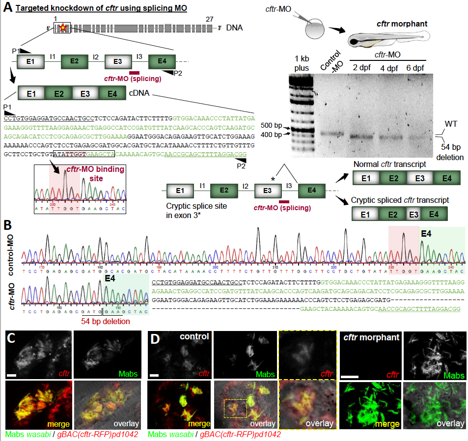
Targeted knockdown of cftr using splicing morpholino in ZF embryos, related to Figure 1 (A-B) A splice targeting morpholino was designed against exon3-intron3 boundary within the cftr gene. (A) Schematic representation and nucleotide sequence corresponding to exon 1 through exon 4 of the ZF cftr gene with the splice morpholino cftr-MO binding site and locations of primers used to amplify the region surrounding exon 3 (P1/P2) (left panel). Altered splicing in cftr MO injected-embryos is verified by RT-PCR (right panel). cftr specific products were amplified from RNA isolated from whole embryos at 2, 4 and 6 dpf. The cftr-MO effectively blocks the splice donor site at the exon3-intron3 boundary, giving rise to an amplicon with a reduce size as compared to the size of the normal cftr transcript, suggesting the existence of a cryptic splice site within exon 3. (B) Comparison of the sequences shows that cftr-MO blocks normal splicing resulting in 54pb deletion in exon 3, thus confirming the efficacy and specificity of this morpholino. (C) Confocal images showing the representative cftr expression in Mabs S- induced granuloma in control-MOinjected gBAC(cftr-RFP)pd104 embryos infected with Mabs expressing Wasabi (5 dpi). Scale bar, 5 μm. (D) Confocal images showing the cftr expression in infected phagocytes in a control or a cftr morphant gBAC(cftr-RFP)pd104 embryos infected with Mabs S expressing Wasabi (5 dpi). Scale bars, 5 μm.