
|
Fig. 3
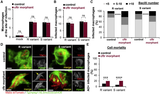
cftr Knockdown Diminishes Intracellular Killing of M. abscessusand Promotes Macrophage Death
(A) Control and cftr morphants Tg(mpeg1:NLSmclover)sh436 were infected with Mabs R or S expressing tdTomato into the hindbrain ventricle (HBV). Confocal microscopy was used to monitor the cell recruitment at 2 hpi. Mean ± SEM number of macrophages recruited to the infected HBV (n = 20, two experiments).
(B) mpeg1:NLSmclover control and cftr morphants were i.v. infected with Mabs R or S expressing tdTomato. Mean ± SEM number of infected macrophages in the caudal hematopoietic tissue (CHT) at 4 hpi (n = 20, two experiments).
(C and D) mpeg1:NLSmclover control and cftr morphants were i.v. infected with Mabs R or S expressing tdTomato imaged at 1 dpi using confocal microscopy to quantify the intracellular bacterial loads.
(C) Average proportions of infected macrophages containing fewer than five, five to ten, or more than ten bacteria in the CHT (n = 16, two experiments).
(D) Confocal images showing infected macrophages. Although WT-macrophages efficiently contain intracellular bacilli, CF macrophages fail to control Mabs growth. Arrow indicates intracellular Mabs R cording. Scale bars, 2 μm.
(E) Control and cftr morphants Tg(mpeg1:mCherry-F)ump2 were i.v. infected with Mabs R or S expressing E2-Crimson and stained with acridine orange (AO). Dead infected macrophages in the CHT were counted using confocal microscopy at 2 dpi. Data are plotted as mean ± SEM from two experiments (n = 20–22).