
|
Fig. S7
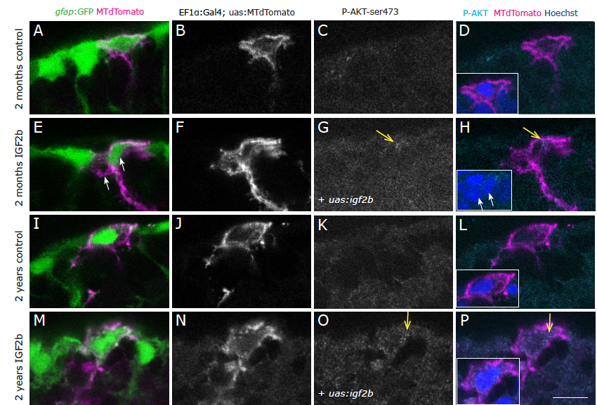
IGF2b overexpression reveals a moderate increase of phospho-Akt immunostaining. Phosphorylated-AKT (Ser473) immunohistochemistry on young (A-H) and old (I-P) brains as a close-up view on the ventricular surface of the pallium. The radial glia somata, located at the border of the ventricle, are labelled in green by the gfap:GFP transgenic line. Single cells stained in magenta have been lipofected in vivo 4 days prior to brain fixation with MTdTomato (control, A-D; I-L) or with MTdTomato and igf2b (IGF2b, E-H; M-P) and are depicted as single confocal plane, highlighting primarily the cell soma (the radial process is not always in the plane of the optical section, therefore not always visible, but it is present below all the somata of lipofected cells). Dots of expression of P-Akt are visible in IGF2b lipofected cells (arrows in G, H, O, P). Insets in D, H, L and P are higher magnifications of the lipofected cells, the nuclei in blue are stained by DAPI. Single confocal planes are displayed, examples are taken out of 3 brains for each condition. Scale bar: 10μm