
|
Fig. S5
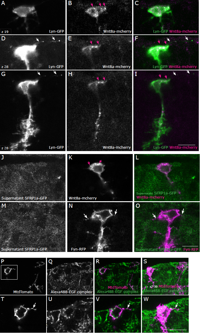
Neither Wnt nor EGF signaling co-localizes with filopodial extensions in adult radial glia. A-I: Example of a cell in a 3-months-old wild-type (AB) brain co-lipofected with pCS2-lyn-GFP and pCS2-wnt8a-mcherry as single confocal planes (z19 in A-C and z28 in D-I). Wnt8a-mcherry localizes in a non- homogeneous manner in the radial glia and can be found at edges of the cytoplasm (pink arrows), however it does not localize in thin filopodial extensions labelled by Lyn-GFP (white arrows). Number of cells analyzed: 8. J-O: The supernatant of Hek-293 cells transfected with sfrp1a-GFP was applied on brain sections containing cells lipofected with pCS2-Wnt8a mcherry (J-L), or with pCS2-fyn-RFP (M-O). The SFRP-GFP protein does not co-localize strongly with Wnt8a-mCherry (pink arrows in K), and does not co-localize with filopodia visible on Fyn-RFP-transfected cells (white arrows in N, O). Number of cells analyzed: 8. P: Staining of a 3-month-old wild-type brain section with the Alexa Fluor 488-EGF complex, which binds to the EGF receptor. Two lipofected cells are visible as a single confocal plane in P-R and as a maximum intensity projection in S. T-W: higher magnification of the inset region depicted in P showing the soma and a filopodial extension (arrow). The EGF-Alexa-488 complex does not co-localize with filopodial extensions. Number of cells analyzed: 12. Scale bars: 10μm.