
|
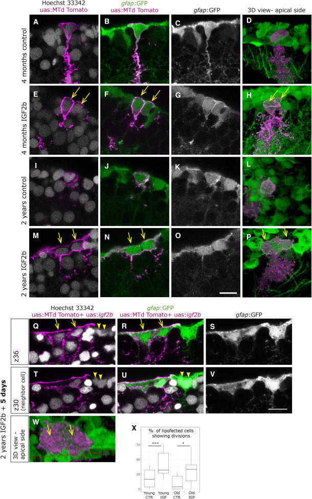
Fig. 6 Overexpression of IFG2b Induces Complete Cell Division in Young Cells, but Incomplete Division in Old Cells (A–P) Brains were lipofected in vivo 4 days prior to fixation with mtdTomato (control, A–D and I–L) or with mtdTomato and igf2b (IGF2b, E–H and M–P). Single confocal planes are displayed in the left panels and 3D reconstructions in the rightmost panels (D, H, L, and P). Cell nuclei (Hoechst, gray) and membrane labeling of the lipofected cells (magenta) are shown in (A), (E), (I), and (M). (A–D) Young control lipofected cells reveal infrequent cell division events, while young cells with IGF2b overexpression (E–H) reveal a higher incidence of cell division events, being visible as two neighboring cells immediately abutting each other (E–H, arrows). (M–P) Old control lipofected cells with IGF2b overexpression result in large cells with two nuclei, indicating an incomplete division (arrows in M, N, and P). (Q–W) Brains fixed 5 days after igf2b lipofection also reveal binucleated large cells (arrows in Q, R, and W), visible here in two distinct confocal planes (Q–S and T–V). Some neighboring cells reveal the same phenotype (arrowheads in Q, T, and U). (X) Cells having divided after lipofection were quantified in 6 experiments with a total of 69 young-control, 114 young-igf2b, 64 old-control, and 71 old-igf2b cells. The significance was calculated by a chi-squared test (young, ∗∗∗p = 0.7 × 10⁻³; old, ∗p = 0.01).