
|
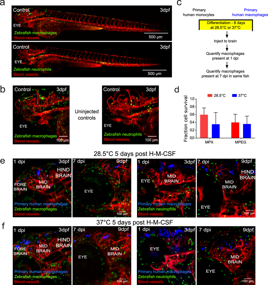
Fig. 6
Primary human macrophages can survive at physiological temperature of the zebrafish in vivo and do not cause a sustained inflammatory response. (a) Representative light sheet micrographs of uninjected, control whole larva at 3 days post-fertilization (dpf). Both transgenic mpeg:GFP (macrophages-green)/flk:mCherry (vessels-red) (top) and mpx:GFP (neutrophils-green)/flk:mCherry (vessels-red) (bottom) are shown. (b) Representative lightsheet micrographs of head regions of uninjected, control larvae at 3 dpf. Both transgenic mpeg:GFP (macrophages-green)/flk:mCherry (vessels-red) (top) and mpx:GFP (neutrophils-green)/flk:mCherry (vessels-red) (bottom) are shown. (c) Schematic of experimental design: primary monocytes were differentiated into macrophages before injection into the zebrafish brain at age 2 dpf and imaged at 1 and 7 days post injection (dpi). (d) Plot (mean ± SD) of average in vivo survival calculated for primary human macrophages differentiated at physiological temperatures of the zebrafish or human obtained from 3 larvae each, where the numbers of cells that survived over the course of 7 days were normalized to the initial numbers measured one day post injection. Differences in survival were not significant by two-way ANOVA with Tukey’s multiple comparisons post-test. The temperature of macrophage differentiation (P = 0.3072), fish line injected (P = 0.4822), and interaction between factors (P = 0.4575) were not significant sources of variation by two-way ANOVA with one degree of freedom. Monocytes that had been cultured in the presence of human macrophage colony stimulating factor (H-M-CSF) for eight days (e) at physiological temperature of (28.5 °C) and (f) at physiological temperature of humans (37 °C). Primary human macrophages (blue) were injected into the hind brain of transgenic mpeg:GFP (macrophages-green)/flk:mCherry (vessels-red) or mpx:GFP (neutrophils-green)/flk:mCherry (vessels-red) zebrafish larvae at 2 dpf. Micrographs of 3D projections showing distribution and survival of human primary macrophages 1 dpi, when larvae are 3 dpf, and 7 dpi, when larvae are 9 dpf. The same fish is shown in the left and right panels.