
|
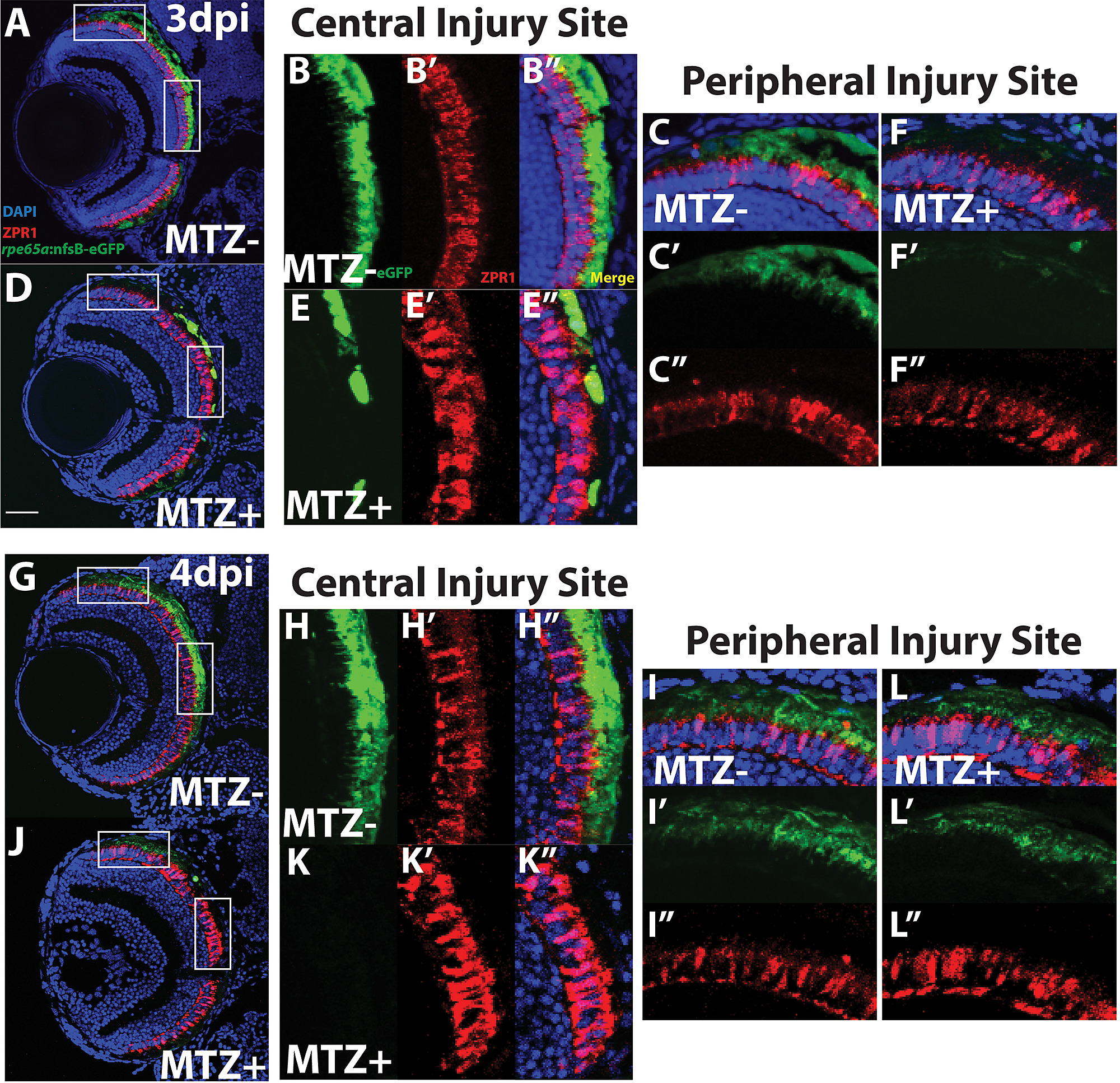
Fig. S4
ZPR1+ PRs adjacent to regenerated RPE in the retinal periphery appear normal.
Transverse cryosections of unablated larvae (A,G) and ablated larvae (D,J) stained for ZPR2. Green = GFP, blue = nuclei, red = ZPR2. Magnified insets of central (B,E) and peripheral (E,F) RPE. (E-E”) At 3dpi, the ZPR1+ signal in PRs within the central injury site becomes less organized and reveals degeneration of outer segment tips. (F-F”) In the periphery, dim GFP signal marks the presence of regenerated RPE, and underlying ZPR1+ cones display morphologies similar to unablated controls. ZPR1+ cones closer to the periphery express normal levels of ZPR1 and identifiable outer segment tips, while those closer to the injury site display increasingly disorganized morphology and lack contiguous outer segment tips. (K-K” At 4dpi, ZPR1 signal continues to be degraded in the central injury site. (L-L”) In the periphery, where GFP+ RPE tissue has regenerated, ZPR1 staining is similar to unablated controls (I-I”), identifying organized cell bodies, outer segments, and outer segment tips. Scale bar = 40μm.