
Fig. S2
- ID
- ZDB-IMAGE-190618-51
- Publication
- Hanovice et al., 2019 - Regeneration of the zebrafish retinal pigment epithelium after widespread genetic ablation
- All Figures
- Figures for Hanovice et al., 2019
|
Fig. S2
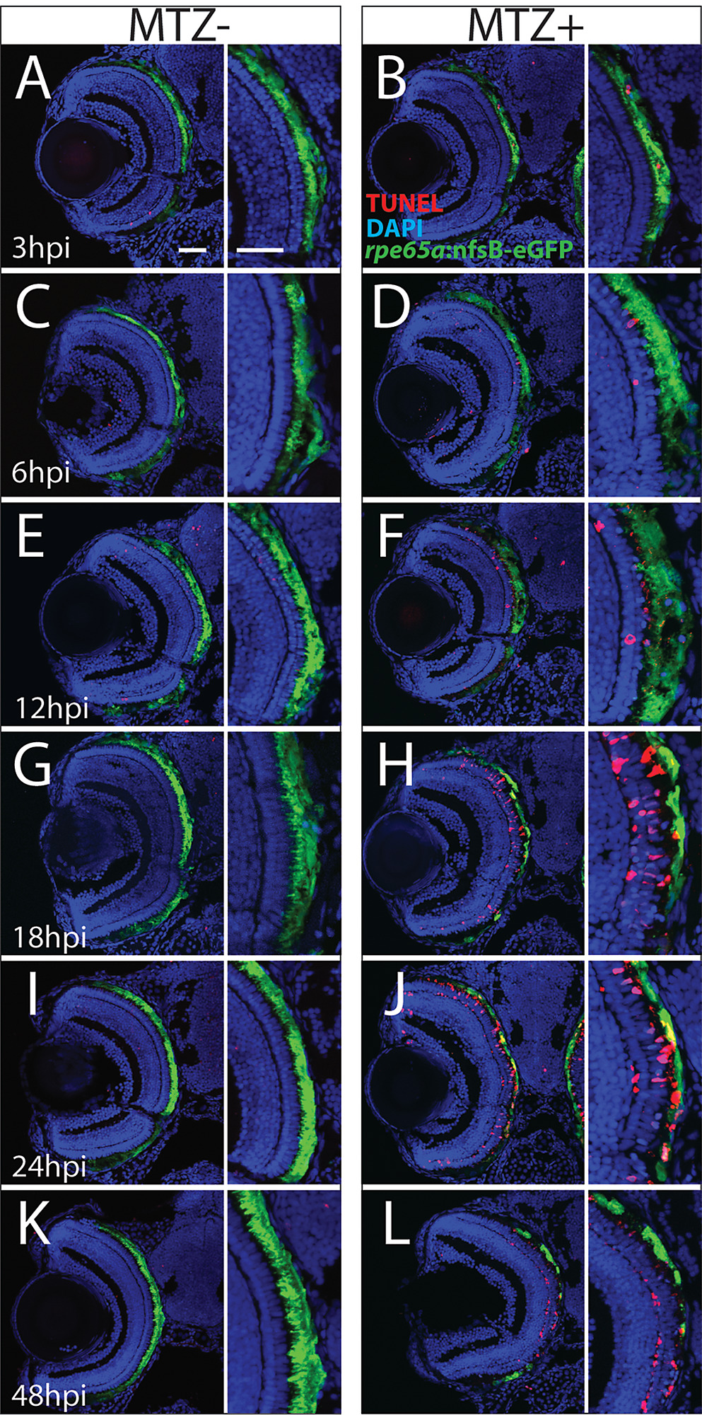
Analysis of TUNEL+ cells in the ablated eye.
(A-L) Transverse cryosections stained for TUNEL (red). (A,C,E,G,I,K) Unablated and (B,D,F,H,J,L) ablated eyes at various time points following ablation. While the ONL appears to be unchanged at 3hpi, slight disruptions in ablated RPE morphology are detectible: apical microvilli become shortened compared to control, and the occasional TUNEL+ nucleus appears in the RPE layer (B). By 6hpi, degeneration of the eGFP+ apical microvilli and cell bodies becomes notable throughout the injury site, and nuclear organization in the ONL begins to degenerate (D). By 18hpi, eGFP signal begins to accumulate in blebs, leaving regions devoid of eGFP+ cells, and TUNEL signal appears throughout the RPE and ONL (H). Degeneration of the central injury site is complete by 48hpi, and TUNEL signal is reduced (L).