
Image
|
Figure Caption
Fig. S1
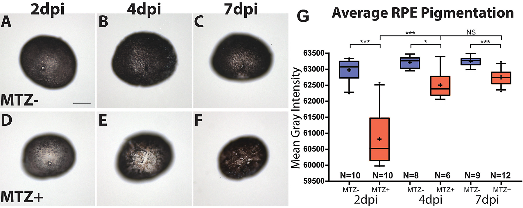
Wholemount analysis of RPE pigmentation.
(A-F) Representative images of wholemounted eyes from unablated (A-C) and ablated larvae (D-F) at 2,4, and 7dpi. (G) Quantification of the mean gray intensity per eye reveals significant depigmentation in ablated larvae at 2dpi, and that pigmentation significantly improves between 2dpi and 4dpi, though overall pigmentation remains significantly reduced compared to unablated controls at 7dpi (Welsh’s t test, * P<0.05, **p<0.005, ***p<0.0005).
Acknowledgments
This image is the copyrighted work of the attributed author or publisher, and
ZFIN has permission only to display this image to its users.
Additional permissions should be obtained from the applicable author or publisher of the image.
Full text @ PLoS Genet.