
|
Fig. 1
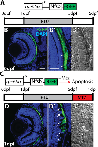
(A) Cartoon depicting the rpe65a:nfsB-eGFP transgene and treatment course of unablated embryos. (B) Transverse cryosections of an unablated 6dpf larva. (B,B’) After exposure to PTU between 1-5dpf, transgene expression is specifically restricted to mature RPE cells, with the brightest expression confined to the central two-thirds of the RPE. Arrowheads indicate apical microvilli. (B”) DIC images reveal RPE repigmentation and normal photoreceptor layer architecture. (C) Cartoon depicting the nitroreductase-mediated ablation paradigm: after washing out PTU, larvae were treated with MTZ for 24 hours. Within cells expressing the transgene, nfsB converts MTZ into a potent DNA crosslinking agent and induces cell death. (D,D’)Transverse cryosections of a 1dpi larva reveal significant disruption of eGFP+ cell morphology and disorganization in INL nuclear lamination. Arrows indicate delaminated and pyknotic nuclei. (D”) DIC images reveal a lack of RPE pigmentation and the marked disruption of photoreceptor layer architecture. Green = eGFP, blue = nuclei. Dorsal is up and distal is left. Scale bar = 40μm.