
Fig. 11
- ID
- ZDB-IMAGE-190618-43
- Antibodies
- Publication
- Hanovice et al., 2019 - Regeneration of the zebrafish retinal pigment epithelium after widespread genetic ablation
- All Figures
- Figures for Hanovice et al., 2019
|
Fig. 11
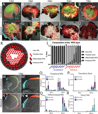
(A-J) en face wholemount images of unablpated (A-E) and ablated (F-J) larvae exposed to 2-hour EdU pulses immediately before fixation and staining for ZPR2 at various time points post-injury. (K) Cartoon depicting the 4 categories into which the RPE was divided, based on cellular pigmentation and intensity of ZPR2 expression: injury site cells peripheral RPE (pigmented, dimly ZPR2+), differentiated RPE (pigmented, ZPR2+), transition zone (lightly pigmented, ZPR2+ consisting of incompletely differentiated RPE extending into the injury site, and injury site (unpigmented, ZPR2-) (L) Quantification of the area of RPE comprised by each domain during regeneration reveals the degeneration of a large proportion of RPE rapidly after ablation, and that regeneration also rapidly occurs, with a transition zone appearing by 1dpi, and differentiated RPE reappearing in the periphery by 2dpi. The entire RPE is repopulated by differentiated RPE by 7dpi. (M-N) Transverse cryosections of unablated (M) and ablated (N) eyes at 1dpi. Magnified insets (M’,N’) reveal the presence of EdU+ cells in the RPE periphery in ablated retinae (N’ arrowhead). (O-R) Quantification of the density of EdU cells throughout regeneration in the peripheral RPE (O), Transition Zone (P), differentiated RPE (Q) and Injury Site (R) suggest that peripheral cells respond to injury by proliferating, that proliferation continues within newly-generated RPE and halts after regeneration is repopulated.