
|
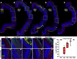
Fig. 9
(A-E) Stitched-together transverse cryosections of unablated (A, n = 4), 3dpi (B, n = 4), 7dpi (C, n = 4), 14dpi (D, n = 3), and 35dpi (E, n = 4) adult eyes, as well as magnified insets of the dorsal peripheral RPE (A’-E’) and central RPE (A”-E”). Red = ZPR2, Green = eGFP, blue = nuclei. At 3dpi, RPE cell degeneration occurred in a large portion of the RPE (B, arrowheads), indicated by loss of eGFP and ZPR2 expression and disruption of overall morphology of the RPE (B’,B”). At 7dpi, increased colocalization of eGFP and ZPR2 defined the peripheral edge of the RPE injury site (C’,C, arrowheads) while the central RPE was absent eGFP and ZPR2 expression (C”). At 14dpi, ZPR2+/eGFP+ RPE reappears in the periphery (D’) and extends inward toward the injury center (D, arrowheads). Magnified images of the central injury site also reveal recovered ZPR2 localization to the apical processes (D”). At 35dpi, ZPR2+/eGFP+ RPE reappear throughout the injury site (E), and proper polarization of ZPR2 and eGFP colocalization has been reestablished throughout (E’-E”). (A-E) Scale bar = 100μm. (A’-E”) Scale bar = 40μm. (F) Quantification of percent RPE regeneration based on measurements of contiguous eGFP+/ZPR2+ expression revealed significant RPE degeneration at 3dpi and 7dpi when compared to MTZ- controls. RPE recovery occurred by 35dpi. Mann-Whitney U Test, * p<0.05.