
Image
|
Figure Caption
Fig. 3
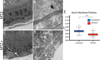
TEM analysis confirms degeneration of the RPE, photoreceptors, and Bruch’s Membrane.
(A,B) TEM images of unablated 8dpf and (C,D) 3dpi eyes. Compared to unablated controls, the ONL and RPE is degenerated in ablated larvae, with large aggregates of debris notable in the RPE (C, arrow). Magnified views of BM reveal reduced BM thickness as well as obvious gaps (D, arrows). (E) Quantification of BM thickness reveals a significant reduction in BM thickness in ablated larvae (Student’s T-test, MTZ- n = 3 eyes, MTZ+ n = 4 eyes p<0.0001).
Figure Data
Acknowledgments
This image is the copyrighted work of the attributed author or publisher, and
ZFIN has permission only to display this image to its users.
Additional permissions should be obtained from the applicable author or publisher of the image.
Full text @ PLoS Genet.