
|
Fig. 3
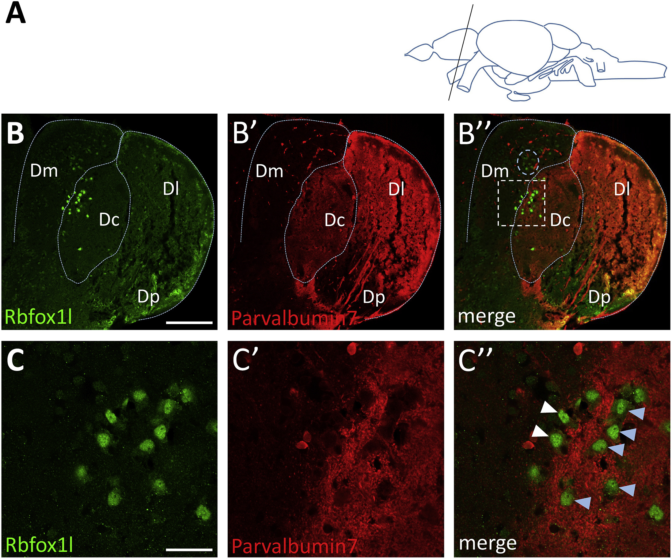
Rbfox1l-positive dorsal telencephalic neurons likely span Dm and Dc regions. (A) Schematic of adult zebrafish brain indicating plane of section through the mid-caudal telencephalon (lateral view). (B-B″) Coronal section of adult zebrafish dorsal telencephalon co-labeled for Parvalbumin7, which labels the Dc and Dl regions (red), and Rbfox1l (green). Dotted blue lines delineating the boundaries of Parvalbuminexpression approximate the telencephalic sub-regions, Dm, Dc, and Dl. Dp region is Parvalbumin7-negative. Rbfox1l-expressing neurons (green) are located largely within the Dc region, but also span the border into the Dm region. A small cluster of Rbfox1l-positive neurons within the Dm are indicated by a dotted blue circle (B″). (C-C″) Higher magnification view of boxed region in (B″). Parvalbumin7 is expressed in many neuronal fibers within the Dc region. Some Rbfox1l-positive neurons are clearly located within the Dc region (light blue arrowheads), while others are located near the border between Dc and Dm (white arrowheads). Abbreviations: Dm, medial zone of the dorsal telencephalon; Dc, central zone of the dorsal telencephalon; Dl, lateral zone of the dorsal telencephalon; Dp, posterior zone of the dorsal telencephalon. Scale bar in B is 150 μm and in C is 30 μm.
Reprinted from Gene expression patterns : GEP, 31, Ma, F., Dong, Z., Berberoglu, M.A., Expression of RNA-binding protein Rbfox1l demarcates a restricted population of dorsal telencephalic neurons within the adult zebrafish brain, 32-41, Copyright (2019) with permission from Elsevier. Full text @ Gene Expr. Patterns