
|
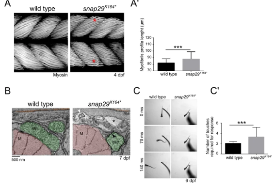
Fig. 4
Snap29 supports correct muscle development and function. (A) Lateral views of muscles in the trunk of 4 dpf wild type and snap29K164* mutant larvae, stained with anti-Myosin heavy chain antibody. Compared to wild type, snap29K164* mutants present less compacted and ordered filaments (red asterisk). (A’) Quantification of the length of the profile of superficial myofibrils measured in wild type and snap29K164* mutant 4 dpf larvae. snap29K164* mutants show a significant increase in myofibril length compared to wild type. The bars in the graph show means and standard deviations. P-values were derived from Mann-Whitney test. ***P ≤ 0.001, n = 41–45. (B) Electron microscopy cross-sections of muscle fibers. snap29K164*contain MLO (white arrowhead) within extracellular spaces (asterisks) just beneath the basement membrane and a mitochondria surrounded by a double membrane (black arrowhead), which are not present in wild type animals. M: myofibrils, Mit: mitochondria, BM: basement membrane. (C) Selected frames from movies of a wild type and a snap29K164*mutant larva at 6 dpf recorded for 1 minute after a touch stimulus on the tail. (C’) Quantification of the number of touches required to evoke an escape response. Most of the analyzed snap29K164* mutants require two or more touches to respond. The graph reports means and standard deviations. P-values were obtained by Mann-Whitney test. ***P ≤ 0.001, n = 6.
v