
|
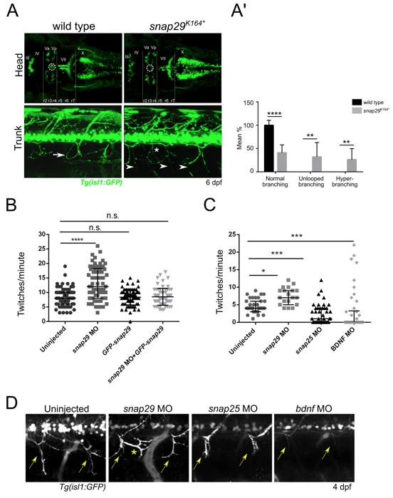
Fig. 5
Snap29 impairment causes neuro-developmental defects. (A, upper row) Head dorsal views of the motor neuron reporter Tg(isl1:GFP) in a wild type and a snap29K164* mutant larva at 6 dpf. (r) rhombomere, (III), (IV), (Va) anterior, (Vp) posterior, (VII), (X) cranial nerves. snap29K164* mutant larvae lack a group of nuclei (white dashed circles) located between the third and fourth rhombomere (r3, r4). (A, lower row) Lateral views of trunks of Tg(isl1:GFP)-expressing wild type and snap29K164* mutants at 6 dpf. Compared to the “loop” structures observed in wild type (white arrow), snap29K164* mutants present an altered motor neuron projection pattern (arrowheads), and extra branching (white asterisk). (A’) Quantification of motor neuron branching in wild type and snap29K164* mutant larvae at 4, 5 and 6 dpf. Compared to wild type, snap29K164* mutants larvae show a significant increase of unlooped branches and hyperbranching. The bars show means and standard deviations of the percentage of the phenotypic categories. P-values were obtained by unpaired t test with Welch’s correction. **P ≤ 0.01, ****P ≤ 0.0001, n = 6–10. (B) Quantification of the number of twitches per minute performed respectively by 26 hpf embryos treated as described in figure. snap29 morphants show an increase in the number of twitches per minute compared to wild type embryos. Co-injection of snap29 Morpholino with GFP-snap29 mRNA rescues the increased number of twitches per minute observed in snap29 morphants, while GFP-snap29 mRNA injection per se has no effect on twitching frequency. The graph shows medians, 25th and 75th percentiles. P-values were obtained by Kruskal-Wallis, ****P ≤ 0.0001, n = 69–73. (C) Quantification of the number of twitches as in B. snap29 morphants show an increase twitches frequency per minute compared to wild type embryos, while both snap25and bdnf morphants show a significant decrease. The graph shows medians, 25th and 75thpercentiles. P-values were derived from Kruskal-Wallis, *P ≤ 0.05 ***P ≤ 0.001, n = 20–44. (D) Motor neuron projections in 4 dpf Tg(isl1:GFP)-expressing uninjected larvae and in snap29, snap25 and bdnf morphant larvae. Uninjected embryos show normal motor neuron projection towards the ventral part of the trunk (yellow arrows). snap29 morphants show less elongated projections (yellow arrows) and extrabranching (yellow asterisk), while both snap25 and bdnf morphants show truncated projections.